 Movie
Movie Controller

[English] 日本語
 Yorodumi
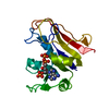
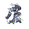
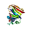
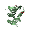
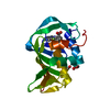
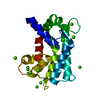
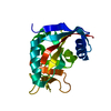
- PDB-5dfr: CRYSTAL STRUCTURE OF UNLIGANDED ESCHERICHIA COLI DIHYDROFOLATE RE... -
+ Open data

- Basic information
| Entry | Database: PDB / ID: 5dfr | ||||||
|---|---|---|---|---|---|---|---|
| Title | CRYSTAL STRUCTURE OF UNLIGANDED ESCHERICHIA COLI DIHYDROFOLATE REDUCTASE. LIGAND-INDUCED CONFORMATIONAL CHANGES AND COOPERATIVITY IN BINDING | ||||||
 Components Components | DIHYDROFOLATE REDUCTASE | ||||||
 Keywords Keywords | OXIDOREDUCTASE / OXIDO-REDUCTASE | ||||||
| Function / homology |  Function and homology informationmethotrexate binding / dihydrofolic acid binding / response to methotrexate / dihydrofolate metabolic process / NADP+ binding / folic acid binding / folic acid metabolic process / glycine biosynthetic process / dihydrofolate reductase / dihydrofolate reductase activity ...methotrexate binding / dihydrofolic acid binding / response to methotrexate / dihydrofolate metabolic process / NADP+ binding / folic acid binding / folic acid metabolic process / glycine biosynthetic process / dihydrofolate reductase / dihydrofolate reductase activity / NADPH binding / tetrahydrofolate biosynthetic process / one-carbon metabolic process / NADP binding / response to xenobiotic stimulus / response to antibiotic / cytosol Function and homology informationmethotrexate binding / dihydrofolic acid binding / response to methotrexate / dihydrofolate metabolic process / NADP+ binding / folic acid binding / folic acid metabolic process / glycine biosynthetic process / dihydrofolate reductase / dihydrofolate reductase activity ...methotrexate binding / dihydrofolic acid binding / response to methotrexate / dihydrofolate metabolic process / NADP+ binding / folic acid binding / folic acid metabolic process / glycine biosynthetic process / dihydrofolate reductase / dihydrofolate reductase activity / NADPH binding / tetrahydrofolate biosynthetic process / one-carbon metabolic process / NADP binding / response to xenobiotic stimulus / response to antibiotic / cytosolSimilarity search - Function | ||||||
| Biological species |  Escherichia coli (E. coli) Escherichia coli (E. coli) | ||||||
| Method | X-RAY DIFFRACTION / Resolution: 2.3 Å | ||||||
 Authors Authors | Bystroff, C. / Kraut, J. | ||||||
 Citation Citation | Journal: Biochemistry / Year: 1991 Title: Crystal structure of unliganded Escherichia coli dihydrofolate reductase. Ligand-induced conformational changes and cooperativity in binding. Authors: Bystroff, C. / Kraut, J. #1: Journal: Biochemistry / Year: 1990Title: Crystal Structures of Escherichia Coli Dihydrofolate Reductase. The Nadp+ Holoenzyme and the Folate(Dot)Nadp+ Ternary Complex. Substrate Binding and a Model for the Transition State Authors: Bystroff, C. / Oatley, S.J. / Kraut, J. #2: Journal: J.Biol.Chem. / Year: 1982Title: Crystal Structures of Escherichia Coli and Lactobacillus Casei Dihydrofolate Reductase Refined at 1.7 Angstroms Resolution. I. General Features and Binding of Methotrexate Authors: Bolin, J.T. / Filman, D.J. / Matthews, D.A. / Hamlin, R.C. / Kraut, J. #3: Journal: Biochemistry / Year: 1987Title: Effect of Single Amino Acid Replacements on the Folding and Stability of Dihydrofolate Reductase from Escherichia Coli Authors: Perry, K.M. / Onuffer, J.J. / Touchette, N.A. / Herndon, C.S. / Gittelman, M.S. / Matthews, C.R. / Chen, J.-T. / Mayer, R.J. / Taira, K. / Benkovic, S.J. / Howell, E.E. / Kraut, J. #4: Journal: J.Biol.Chem. / Year: 1982Title: Crystal Structures of Escherichia Coli and Lactobacillus Casei Dihydrofolate Reductase Refined at 1.7 Angstroms Resolution. II. Environment of Bound Nadph and Implications for Catalysis Authors: Filman, D.J. / Bolin, J.T. / Matthews, D.A. / Kraut, J. #5: Journal: J.Biol.Chem. / Year: 1982Title: Crystal Structure of Avian Dihydrofolate Reductase Containing Phenyltriazine and Nadph Authors: Volz, K.W. / Matthews, D.A. / Alden, R.A. / Freer, S.T. / Hansch, C. / Kaufman, B.T. / Kraut, J. #6: Journal: Biochemistry / Year: 1979Title: Interpretation of Nuclear Magnetic Resonance Spectra for Lactobacillus Casei Dihydrofolate Reductase Based on the X-Ray Structure of the Enzyme-Methotrexate-Nadph Complex Authors: Matthews, D.A. #7: Journal: J.Biol.Chem. / Year: 1979Title: Dihydrofolate Reductase from Lactobacillus Casei. Stereochemistry of Nadph Binding Authors: Matthews, D.A. / Alden, R.A. / Freer, S.T. / Xuong, N.-H. / Kraut, J. #8: Journal: J.Biol.Chem. / Year: 1979Title: Proton Magnetic Resonance Studies on Escherichia Coli Dihydrofolate Reductase. Assignment of Histidine C-2 Protons in Binary Complexes with Folates on the Basis of the Crystal Structure with ...Title: Proton Magnetic Resonance Studies on Escherichia Coli Dihydrofolate Reductase. Assignment of Histidine C-2 Protons in Binary Complexes with Folates on the Basis of the Crystal Structure with Methotrexate and on Chemical Modifications Authors: Poe, M. / Hoogsteen, K. / Matthews, D.A. #9: Journal: J.Biol.Chem. / Year: 1978Title: Dihydrofolate Reductase from Lactobacillus Casei. X-Ray Structure of the Enzyme-Methotrexate-Nadph Complex Authors: Matthews, D.A. / Alden, R.A. / Bolin, J.T. / Filman, D.J. / Freer, S.T. / Hamlin, R. / Hol, W.G.J. / Kisliuk, R.L. / Pastore, E.J. / Plante, L.T. / Xuong, N.-H. / Kraut, J. #10: Journal: Biochemistry / Year: 1978Title: Dihydrofolate Reductase. The Amino Acid Sequence of the Enzyme from a Methotrexate-Resistant Mutant of Escherichia Coli Authors: Bennett, C.D. / Rodkey, J.A. / Sondey, J.M. / Hirschmann, R. #11: Journal: Science / Year: 1977Title: Dihydrofolate Reductase. X-Ray Structure of the Binary Complex with Methotrexate Authors: Matthews, D.A. / Alden, R.A. / Bolin, J.T. / Freer, S.T. / Hamlin, R. / Xuong, N. / Kraut, J. / Poe, M. / Williams, M. / Hoogsteen, K. #12: Journal: Biochemistry / Year: 1972Title: Dihydrofolate Reductase. Purification and Characterization of the Enzyme from an Amethopterin-Resistant Mutant of Escherichia Coli Authors: Poe, M. / Greenfield, N.J. / Hirshfield, J.M. / Williams, M.N. / Hoogsteen, K. | ||||||
| History |
|
- Structure visualization
| Structure viewer | Molecule: MolmilJmol/JSmol |
|---|
- Downloads & links
-Download
| PDBx/mmCIF format | 5dfr.cif.gz | 48.3 KB | Display | PDBx/mmCIF format |
|---|---|---|---|---|
| PDB format | pdb5dfr.ent.gz | 34.1 KB | Display | PDB format |
| PDBx/mmJSON format | 5dfr.json.gz | Tree view | PDBx/mmJSON format | |
| Others |  Other downloads Other downloads |
-Validation report
| Arichive directory | https://data.pdbj.org/pub/pdb/validation_reports/df/5dfrftp://data.pdbj.org/pub/pdb/validation_reports/df/5dfr | HTTPS FTP |
|---|
-Related structure data


-Links
 PDBj
PDBj
- Assembly
| Deposited unit | 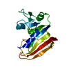
| ||||||||
|---|---|---|---|---|---|---|---|---|---|
| 1 |
| ||||||||
| Unit cell |
| ||||||||
| Atom site foot note | 1: RESIDUES PRO 21 - LEU 24 ARE WEAK. THERE IS UNINTERPRETED DENSITY ASSOCIATED WITH THE SIDE CHAIN OF PHE 31. RESIDUES 95 - 96 ARE PARTIALLY DISORDERED. VAL 10, GLU 120, ASP 122, AND ASP 127 HAVE ...1: RESIDUES PRO 21 - LEU 24 ARE WEAK. THERE IS UNINTERPRETED DENSITY ASSOCIATED WITH THE SIDE CHAIN OF PHE 31. RESIDUES 95 - 96 ARE PARTIALLY DISORDERED. VAL 10, GLU 120, ASP 122, AND ASP 127 HAVE DISORDERED SIDE CHAINS. THE SIDE CHAIN OF LEU 28, IN THE SUBSTRATE BINDING SITE, APPEARS TO BE PARTIALLY DISORDERED. 2: THE IDENTITY OF IONS 501 THROUGH 503 IS NOT DETERMINED. THEY HAVE TENATIVELY BEEN ASSIGNED AS CHLORINE IONS BASED ON THEIR ELECTRON DENSITY, THE BINDING ENVIRONMENT, AND THE BELIEF THAT CHLORIDE ...2: THE IDENTITY OF IONS 501 THROUGH 503 IS NOT DETERMINED. THEY HAVE TENATIVELY BEEN ASSIGNED AS CHLORINE IONS BASED ON THEIR ELECTRON DENSITY, THE BINDING ENVIRONMENT, AND THE BELIEF THAT CHLORIDE IS THE ONLY NEGATIVELY-CHARGED MONO-ATOMIC ION PRESENT IN THE CRYSTALLIZATION MIXTURE. |
-Components
| #1: Protein | Mass: 18020.326 Da / Num. of mol.: 1 Source method: isolated from a genetically manipulated source Source: (gene. exp.) Escherichia coli (E. coli) / References: UniProt: P0ABQ4, dihydrofolate reductase | ||||
|---|---|---|---|---|---|
| #2: Chemical | Chloride  Mass: 35.453 Da / Num. of mol.: 3 / Source method: obtained synthetically / Formula: Cl Mass: 35.453 Da / Num. of mol.: 3 / Source method: obtained synthetically / Formula: Cl#3: Water | ChemComp-HOH / | Water Mass: 18.015 Da / Num. of mol.: 120 / Source method: isolated from a natural source / Formula: H2O Mass: 18.015 Da / Num. of mol.: 120 / Source method: isolated from a natural source / Formula: H2ONonpolymer details | THE IDENTITY OF IONS 501 THROUGH 503 IS NOT DETERMINED. THEY HAVE TENATIVELY BEEN ASSIGNED AS ...THE IDENTITY OF IONS 501 THROUGH 503 IS NOT DETERMINED | |
-Experimental details
-Experiment
| Experiment | Method: X-RAY DIFFRACTION |
|---|
- Sample preparation
| Crystal | Density Matthews: 3.15 Å3/Da / Density % sol: 60.98 % | ||||||||||||||||||||||||||||||
|---|---|---|---|---|---|---|---|---|---|---|---|---|---|---|---|---|---|---|---|---|---|---|---|---|---|---|---|---|---|---|---|
| Crystal grow | *PLUS Temperature: 4 ℃ / pH: 5.4 / Method: vapor diffusion, hanging drop | ||||||||||||||||||||||||||||||
| Components of the solutions | *PLUS
|
-Data collection
| Reflection | *PLUS Highest resolution: 2.05 Å / Lowest resolution: 2.3 Å / Num. all: 63697 / Num. obs: 14169 / Num. measured all: 14730 / Rmerge(I) obs: 0.043 |
|---|
- Processing
| Software | Name: PROLSQ / Classification: refinement | ||||||||||||||||||||||||||||||||||||||||||||||||||||||||||||||||||||||||||||||||||||
|---|---|---|---|---|---|---|---|---|---|---|---|---|---|---|---|---|---|---|---|---|---|---|---|---|---|---|---|---|---|---|---|---|---|---|---|---|---|---|---|---|---|---|---|---|---|---|---|---|---|---|---|---|---|---|---|---|---|---|---|---|---|---|---|---|---|---|---|---|---|---|---|---|---|---|---|---|---|---|---|---|---|---|---|---|---|
| Refinement | Resolution: 2.3→20 Å / Rfactor obs: 0.198 Details: THE PAPER CITED ON THE *JRNL* RECORDS ABOVE DISCUSSES THE QUESTION AS TO WHETHER THE PEPTIDE BOND BETWEEN GLY 95 AND GLY 96 IS IN THE CIS OR THE TRANS CONFORMATION. A DISORDERED MODEL IN ...Details: THE PAPER CITED ON THE *JRNL* RECORDS ABOVE DISCUSSES THE QUESTION AS TO WHETHER THE PEPTIDE BOND BETWEEN GLY 95 AND GLY 96 IS IN THE CIS OR THE TRANS CONFORMATION. A DISORDERED MODEL IN WHICH THIS PEPTIDE EXISTS IN BOTH CONFORMERS CANNOT BE RULED OUT. IN THIS ENTRY, HOWEVER, ONLY COORDINATES CORRESPONDING TO THE TRANS CONFORMATION ARE PROVIDED. RESIDUES PRO 21 - LEU 24 ARE WEAK. THERE IS UNINTERPRETED DENSITY ASSOCIATED WITH THE SIDE CHAIN OF PHE 31. RESIDUES GLY 95 - GLY 96 ARE PARTIALLY DISORDERED. VAL 10, GLU 120, ASP 122, AND ASP 127 HAVE DISORDERED SIDE CHAINS. THE SIDE CHAIN OF LEU 28, IN THE SUBSTRATE BINDING SITE, APPEARS TO BE PARTIALLY DISORDERED. | ||||||||||||||||||||||||||||||||||||||||||||||||||||||||||||||||||||||||||||||||||||
| Refinement step | Cycle: LAST / Resolution: 2.3→20 Å
| ||||||||||||||||||||||||||||||||||||||||||||||||||||||||||||||||||||||||||||||||||||
| Refine LS restraints |
| ||||||||||||||||||||||||||||||||||||||||||||||||||||||||||||||||||||||||||||||||||||
| Refinement | *PLUS Highest resolution: 2.3 Å / Lowest resolution: 20 Å / Rfactor obs: 0.198 | ||||||||||||||||||||||||||||||||||||||||||||||||||||||||||||||||||||||||||||||||||||
| Solvent computation | *PLUS | ||||||||||||||||||||||||||||||||||||||||||||||||||||||||||||||||||||||||||||||||||||
| Displacement parameters | *PLUS Biso mean: 31 Å2 |
