Mass: 18.015 Da / Num. of mol.: 21 / Source method: isolated from a natural source / Formula: H2O
Sequence details
1. SEQUENCE CONFLICTS D270G, A337T AND I479V ARE BASED ON BAB43823.1 ACCORDING TO DATABASE Q9C0L9 ...1. SEQUENCE CONFLICTS D270G, A337T AND I479V ARE BASED ON BAB43823.1 ACCORDING TO DATABASE Q9C0L9 (SEY1_CANAL). 2. THE CODON CUG (MRNA CUG CORRESPONDS TO CTG IN DNA) WILL ENCODE SER IN CANDIDA ALBICANS BUT LEU IN ESCHERICHIA COLI. SO 89S,221S,665S IN CANDIDA ALBICANS SEY1P(BAB43817) WILL BE TRANSLATED INTO LEU IN ESCHERICHIA COLI BL21.
-
Experimental details
-
Experiment
Experiment
Method: X-RAY DIFFRACTION / Number of used crystals: 1
-
Sample preparation
Crystal
Density Matthews: 2.79 Å3/Da / Density % sol: 55.85 %
Crystal grow
Temperature: 289 K / Method: evaporation Details: 120 Mm DL-Malic acid pH 7.0, 16% w/v PEG 3350, 20 mM Bicine pH 9.0, 400 mM Magnesium chloride hexahydrate, 6% D-(+)-Trehalose dihydrate
Protocol: SINGLE WAVELENGTH / Monochromatic (M) / Laue (L): M / Scattering type: x-ray
Radiation wavelength
Wavelength: 1 Å / Relative weight: 1
Reflection
Resolution: 2.8→50 Å / Num. obs: 19897 / % possible obs: 99.5 % / Redundancy: 6.9 % / Net I/σ(I): 27.2
-
Processing
Software
Name
Version
Classification
REFMAC
5.8.0049
refinement
HKL-2000
dataprocessing
Coot
modelbuilding
Refinement
Resolution: 2.9→50 Å / Cor.coef. Fo:Fc: 0.909 / Cor.coef. Fo:Fc free: 0.897 / SU B: 45.901 / SU ML: 0.412 / Cross valid method: THROUGHOUT / ESU R Free: 0.469 / Stereochemistry target values: MAXIMUM LIKELIHOOD / Details: HYDROGENS HAVE BEEN ADDED IN THE RIDING POSITIONS
Rfactor
Num. reflection
% reflection
Selection details
Rfree
0.29806
1015
5.1 %
RANDOM
Rwork
0.2596
-
-
-
obs
0.26156
18876
99.37 %
-
Solvent computation
Ion probe radii: 0.8 Å / Shrinkage radii: 0.8 Å / VDW probe radii: 1.2 Å / Solvent model: MASK
 Movie
Movie Controller


 Open data
 Basic information
 Components
 Keywords
 Function and homology information
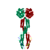
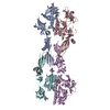
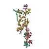
 Candida albicans (yeast)
 X-RAY DIFFRACTION /
  Authors
 China, 3items
  Citation
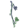
 Structure visualization
 Downloads & links
 Other downloads
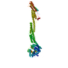

 PDBj
PDBj


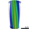
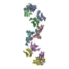
 Assembly
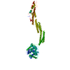


 Mass: 24.305 Da / Num. of mol.: 1 / Source method: obtained synthetically / Formula: Mg
 Mass: 522.196 Da / Num. of mol.: 1 / Source method: obtained synthetically / Formula: C10H17N6O13P3
 Mass: 18.015 Da / Num. of mol.: 21 / Source method: isolated from a natural source / Formula: H2O
 Sample preparation
 Processing