[English] 日本語
Yorodumi
- PDB-5ca8: Structures of the yeast dynamin-like GTPase Sey1p in complex with GDP -

+
Open data


ID or keywords:

Loading...

-
Basic information

Entry
Database: PDB / ID: 5ca8
TitleStructures of the yeast dynamin-like GTPase Sey1p in complex with GDP
ComponentsProtein SEY1
KeywordsHYDROLASE / ER / Homo-fusion / Dynamin
Function / homology
Function and homology information


endoplasmic reticulum inheritance / hexose transmembrane transport / endoplasmic reticulum membrane fusion / cortical endoplasmic reticulum / Hydrolases; Acting on acid anhydrides; Acting on GTP to facilitate cellular and subcellular movement / GTPase activity / endoplasmic reticulum membrane / GTP binding / endoplasmic reticulum
Similarity search - Function
RHD3/Sey1 / Sey1/RHD3-like, three-helix bundle domain / Root hair defective 3 GTP-binding protein (RHD3) GTPase domain / Sey1 three-helix bundle domain / GB1/RHD3-type guanine nucleotide-binding (G) domain / GB1/RHD3-type guanine nucleotide-binding (G) domain profile. / P-loop containing nucleoside triphosphate hydrolase
Similarity search - Domain/homology
GUANOSINE-5'-DIPHOSPHATE / Protein SEY1
Similarity search - Component
Biological species
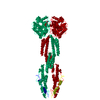
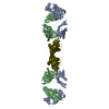
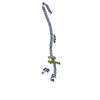
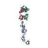
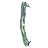
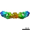
Candida albicans (yeast)
MethodX-RAY DIFFRACTION / SYNCHROTRON / MOLECULAR REPLACEMENT / Resolution: 2.3 Å
AuthorsYan, L.
Funding support China, 2items
OrganizationGrant numberCountry
National Natural Science Foundation81322023 China
973 Program2013CB911103 China
CitationJournal: J.Cell Biol. / Year: 2015
Title: Structures of the yeast dynamin-like GTPase Sey1p provide insight into homotypic ER fusion
Authors: Yan, L. / Sun, S. / Wang, W. / Shi, J. / Hu, X. / Wang, S. / Su, D. / Rao, Z. / Hu, J. / Lou, Z.
History
DepositionJun 29, 2015Deposition site: RCSB / Processing site: PDBJ
Revision 1.0Sep 16, 2015Provider: repository / Type: Initial release
Revision 1.1Oct 7, 2015Group: Database references
Revision 1.2Feb 19, 2020Group: Data collection / Database references / Derived calculations
Category: citation / diffrn_source / pdbx_struct_oper_list
Item: _citation.journal_id_CSD / _diffrn_source.pdbx_synchrotron_site / _pdbx_struct_oper_list.symmetry_operation
Revision 1.3Nov 8, 2023Group: Data collection / Database references ...Data collection / Database references / Derived calculations / Refinement description
Category: chem_comp_atom / chem_comp_bond ...chem_comp_atom / chem_comp_bond / database_2 / pdbx_initial_refinement_model / pdbx_struct_conn_angle / struct_conn / struct_conn_type
Item: _database_2.pdbx_DOI / _database_2.pdbx_database_accession ..._database_2.pdbx_DOI / _database_2.pdbx_database_accession / _pdbx_struct_conn_angle.ptnr1_auth_comp_id / _pdbx_struct_conn_angle.ptnr1_auth_seq_id / _pdbx_struct_conn_angle.ptnr1_label_asym_id / _pdbx_struct_conn_angle.ptnr1_label_atom_id / _pdbx_struct_conn_angle.ptnr1_label_comp_id / _pdbx_struct_conn_angle.ptnr3_auth_comp_id / _pdbx_struct_conn_angle.ptnr3_auth_seq_id / _pdbx_struct_conn_angle.ptnr3_label_asym_id / _pdbx_struct_conn_angle.ptnr3_label_atom_id / _pdbx_struct_conn_angle.ptnr3_label_comp_id / _pdbx_struct_conn_angle.value / _struct_conn.conn_type_id / _struct_conn.id / _struct_conn.pdbx_dist_value / _struct_conn.pdbx_leaving_atom_flag / _struct_conn.ptnr1_auth_comp_id / _struct_conn.ptnr1_auth_seq_id / _struct_conn.ptnr1_label_atom_id / _struct_conn.ptnr1_label_comp_id / _struct_conn.ptnr1_label_seq_id / _struct_conn.ptnr2_auth_comp_id / _struct_conn.ptnr2_auth_seq_id / _struct_conn.ptnr2_label_asym_id / _struct_conn.ptnr2_label_atom_id / _struct_conn.ptnr2_label_comp_id / _struct_conn.ptnr2_label_seq_id / _struct_conn_type.id

-
Structure visualization

Structure viewerMolecule:
MolmilJmol/JSmol

Downloads & links

-
Assembly

Deposited unit
A: Protein SEY1
hetero molecules


Theoretical massNumber of molelcules
Total (without water)79,5923
Polymers79,1241
Non-polymers4682
Water2,432135
1


  • Idetical with deposited unit
  • defined by author&software
TypeNameSymmetry operationNumber
identity operation1_555x,y,z1
Buried area850 Å2
ΔGint-16 kcal/mol
Surface area32990 Å2
MethodPISA
Unit cell
Length a, b, c (Å)228.120, 43.620, 97.560
Angle α, β, γ (deg.)90.00, 108.12, 90.00
Int Tables number5
Space group name H-MC121
Components on special symmetry positions
IDModelComponents
11A-801-

HOH

-
Components

#1: Protein Protein SEY1


Mass: 79124.000 Da / Num. of mol.: 1 / Fragment: UNP residues 1-692
Source method: isolated from a genetically manipulated source
Source: (gene. exp.) Candida albicans (strain SC5314 / ATCC MYA-2876) (yeast)
Strain: SC5314 / ATCC MYA-2876 / Gene: SEY1, NAG6, CaO19.2151, CaO19.9698 / Production host: Escherichia coli (E. coli) / References: UniProt: Q9C0L9, dynamin GTPase
#2: Chemical ChemComp-MG / MAGNESIUM ION


Mass: 24.305 Da / Num. of mol.: 1 / Source method: obtained synthetically / Formula: Mg
#3: Chemical ChemComp-GDP / GUANOSINE-5'-DIPHOSPHATE


Type: RNA linking / Mass: 443.201 Da / Num. of mol.: 1 / Source method: obtained synthetically / Formula: C10H15N5O11P2 / Comment: GDP, energy-carrying molecule*YM
#4: Water ChemComp-HOH / water


Mass: 18.015 Da / Num. of mol.: 135 / Source method: isolated from a natural source / Formula: H2O
Sequence details1. SEQUENCE CONFLICTS D270G, A337T AND I479V ARE BASED ON BAB43823.1 ACCORDING TO DATABASE Q9C0L9 ...1. SEQUENCE CONFLICTS D270G, A337T AND I479V ARE BASED ON BAB43823.1 ACCORDING TO DATABASE Q9C0L9 (SEY1_CANAL). 2. THE CODON CUG (MRNA CUG CORRESPONDS TO CTG IN DNA) WILL ENCODE SER IN CANDIDA ALBICANS BUT LEU IN ESCHERICHIA COLI. SO 89S,221S,665S IN CANDIDA ALBICANS SEY1P(BAB43817) WILL BE TRANSLATED INTO LEU IN ESCHERICHIA COLI BL21.

-
Experimental details

-
Experiment

ExperimentMethod: X-RAY DIFFRACTION / Number of used crystals: 1

-
Sample preparation

CrystalDensity Matthews: 2.92 Å3/Da / Density % sol: 57.81 %
Crystal growTemperature: 289 K / Method: evaporation
Details: 20 mM Tris pH 8.5, 160 mM Potassium sodium tartrate tetrahydrate, 4 mM GDP, 21% w/v PEG3350

-
Data collection

DiffractionMean temperature: 100 K
Diffraction sourceSource: SYNCHROTRON / Site: Diamond / Beamline: I03 / Wavelength: 0.9795 Å
DetectorType: PSI PILATUS 6M / Detector: PIXEL / Date: Sep 23, 2013
RadiationProtocol: SINGLE WAVELENGTH / Monochromatic (M) / Laue (L): M / Scattering type: x-ray
Radiation wavelengthWavelength: 0.9795 Å / Relative weight: 1
ReflectionResolution: 2.3→50 Å / Num. obs: 40802 / % possible obs: 99.1 % / Redundancy: 2.99 % / Net I/σ(I): 10.2

-
Processing

Software
NameVersionClassification
PHENIX1.8.4_1496refinement
HKL-2000data processing
PHENIXmodel building
PHASERphasing
RefinementMethod to determine structure: MOLECULAR REPLACEMENT
Starting model: 5CA9
Resolution: 2.3→46.361 Å / SU ML: 0.38 / Cross valid method: THROUGHOUT / σ(F): 1.36 / Phase error: 31.42 / Stereochemistry target values: ML
RfactorNum. reflection% reflectionSelection details
Rfree0.285 2049 5.02 %RANDOM
Rwork0.2304 ---
obs0.2331 40796 98.89 %-
Solvent computationShrinkage radii: 0.9 Å / VDW probe radii: 1.11 Å / Solvent model: FLAT BULK SOLVENT MODEL
Refinement stepCycle: LAST / Resolution: 2.3→46.361 Å
ProteinNucleic acidLigandSolventTotal
Num. atoms5136 0 29 135 5300
Refine LS restraints
Refine-IDTypeDev idealNumber
X-RAY DIFFRACTIONf_bond_d0.0115288
X-RAY DIFFRACTIONf_angle_d1.5147150
X-RAY DIFFRACTIONf_dihedral_angle_d18.21945
X-RAY DIFFRACTIONf_chiral_restr0.063810
X-RAY DIFFRACTIONf_plane_restr0.007913
LS refinement shell
Resolution (Å)Rfactor RfreeNum. reflection RfreeRfactor RworkNum. reflection RworkRefine-ID% reflection obs (%)
2.3-2.35350.37781490.34612524X-RAY DIFFRACTION99
2.3535-2.41240.34951280.3042588X-RAY DIFFRACTION99
2.4124-2.47760.33641520.29552519X-RAY DIFFRACTION98
2.4776-2.55050.31951280.27052592X-RAY DIFFRACTION99
2.5505-2.63280.31221460.26362536X-RAY DIFFRACTION99
2.6328-2.72690.30921450.25152568X-RAY DIFFRACTION98
2.7269-2.83610.30571080.2482523X-RAY DIFFRACTION98
2.8361-2.96510.29891460.2422575X-RAY DIFFRACTION98
2.9651-3.12140.29931150.23642598X-RAY DIFFRACTION98
3.1214-3.31690.25861220.2412577X-RAY DIFFRACTION99
3.3169-3.5730.28381460.21582572X-RAY DIFFRACTION99
3.573-3.93240.26351470.20772582X-RAY DIFFRACTION98
3.9324-4.5010.26011310.19292596X-RAY DIFFRACTION99
4.501-5.66910.27551380.20282644X-RAY DIFFRACTION99
5.6691-46.37020.28441480.24662753X-RAY DIFFRACTION100
Refinement TLS params.Method: refined / Origin x: 36.9668 Å / Origin y: 7.1418 Å / Origin z: 6.4301 Å
111213212223313233
T0.4719 Å20.0622 Å2-0.1058 Å2-0.6169 Å20.0362 Å2--0.5813 Å2
L0.7904 °2-0.3542 °20.8358 °2-0.0296 °2-0.2905 °2--0.5309 °2
S0.0525 Å °-0.1114 Å °-0.0856 Å °-0.0884 Å °0.0221 Å °0.0445 Å °0.0547 Å °-0.2048 Å °-0.0237 Å °
Refinement TLS groupSelection details: all

+
About Yorodumi

-
News

-
Feb 9, 2022. New format data for meta-information of EMDB entries

New format data for meta-information of EMDB entries

  • Version 3 of the EMDB header file is now the official format.
  • The previous official version 1.9 will be removed from the archive.

Related info.:EMDB header

External links:wwPDB to switch to version 3 of the EMDB data model

-
Aug 12, 2020. Covid-19 info

Covid-19 info

URL: https://pdbjlvh1.pdbj.org/emnavi/covid19.php

New page: Covid-19 featured information page in EM Navigator.

Related info.:Covid-19 info / Mar 5, 2020. Novel coronavirus structure data

+
Mar 5, 2020. Novel coronavirus structure data

Novel coronavirus structure data

Related info.:Yorodumi Speices / Aug 12, 2020. Covid-19 info

External links:COVID-19 featured content - PDBj / Molecule of the Month (242):Coronavirus Proteases

+
Jan 31, 2019. EMDB accession codes are about to change! (news from PDBe EMDB page)

EMDB accession codes are about to change! (news from PDBe EMDB page)

  • The allocation of 4 digits for EMDB accession codes will soon come to an end. Whilst these codes will remain in use, new EMDB accession codes will include an additional digit and will expand incrementally as the available range of codes is exhausted. The current 4-digit format prefixed with “EMD-” (i.e. EMD-XXXX) will advance to a 5-digit format (i.e. EMD-XXXXX), and so on. It is currently estimated that the 4-digit codes will be depleted around Spring 2019, at which point the 5-digit format will come into force.
  • The EM Navigator/Yorodumi systems omit the EMD- prefix.

Related info.:Q: What is EMD? / ID/Accession-code notation in Yorodumi/EM Navigator

External links:EMDB Accession Codes are Changing Soon! / Contact to PDBj

+
Jul 12, 2017. Major update of PDB

Major update of PDB

  • wwPDB released updated PDB data conforming to the new PDBx/mmCIF dictionary.
  • This is a major update changing the version number from 4 to 5, and with Remediation, in which all the entries are updated.
  • In this update, many items about electron microscopy experimental information are reorganized (e.g. em_software).
  • Now, EM Navigator and Yorodumi are based on the updated data.

External links:wwPDB Remediation / Enriched Model Files Conforming to OneDep Data Standards Now Available in the PDB FTP Archive

-
Yorodumi

Thousand views of thousand structures

  • Yorodumi is a browser for structure data from EMDB, PDB, SASBDB, etc.
  • This page is also the successor to EM Navigator detail page, and also detail information page/front-end page for Omokage search.
  • The word "yorodu" (or yorozu) is an old Japanese word meaning "ten thousand". "mi" (miru) is to see.

Related info.:EMDB / PDB / SASBDB / Comparison of 3 databanks / Yorodumi Search / Aug 31, 2016. New EM Navigator & Yorodumi / Yorodumi Papers / Jmol/JSmol / Function and homology information / Changes in new EM Navigator and Yorodumi

Read more