 Movie
Movie Controller


+ Open data

- Basic information
| Entry | Database: PDB / ID: 5c8h | ||||||
|---|---|---|---|---|---|---|---|
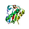
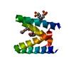
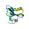
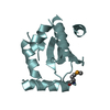
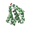
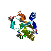
| Title | Crystal structure of ORC2 C-terminal domain | ||||||
 Components Components | Origin recognition complex subunit 2 | ||||||
 Keywords Keywords | REPLICATION / Structural Genomics / DNA replication / Structural Genomics Consortium / SGC | ||||||
| Function / homology |  Function and homology information Function and homology informationCDC6 association with the ORC:origin complex / origin recognition complex / E2F-enabled inhibition of pre-replication complex formation / nuclear origin of replication recognition complex / inner kinetochore / DNA replication origin binding / DNA replication initiation / Activation of the pre-replicative complex / Activation of ATR in response to replication stress / heterochromatin ...CDC6 association with the ORC:origin complex / origin recognition complex / E2F-enabled inhibition of pre-replication complex formation / nuclear origin of replication recognition complex / inner kinetochore / DNA replication origin binding / DNA replication initiation / Activation of the pre-replicative complex / Activation of ATR in response to replication stress / heterochromatin / Assembly of the ORC complex at the origin of replication / Assembly of the pre-replicative complex / Orc1 removal from chromatin / chromosome, telomeric region / centrosome / negative regulation of transcription by RNA polymerase II / nucleoplasm / membrane / nucleus Similarity search - Function | ||||||
| Biological species |  Homo sapiens (human) Homo sapiens (human) | ||||||
| Method |  X-RAY DIFFRACTION / SYNCHROTRON / MOLECULAR REPLACEMENT / molecular replacement / Resolution: 2.01 Å X-RAY DIFFRACTION / SYNCHROTRON / MOLECULAR REPLACEMENT / molecular replacement / Resolution: 2.01 Å | ||||||
 Authors Authors | Tempel, W. / Xu, C. / Dong, A. / Loppnau, P. / Bountra, C. / Arrowsmith, C.H. / Edwards, A.M. / Min, J. / Structural Genomics Consortium (SGC) | ||||||
 Citation Citation | Journal: To Be Published Title: Crystal structure of ORC2 C-terminal domain Authors: Tempel, W. / Xu, C. / Dong, A. / Loppnau, P. / Bountra, C. / Arrowsmith, C.H. / Edwards, A.M. / Min, J. | ||||||
| History |
|
- Structure visualization
| Structure viewer | Molecule: MolmilJmol/JSmol |
|---|
- Downloads & links
-Download
| PDBx/mmCIF format | 5c8h.cif.gz | 57.7 KB | Display | PDBx/mmCIF format |
|---|---|---|---|---|
| PDB format | pdb5c8h.ent.gz | 40.7 KB | Display | PDB format |
| PDBx/mmJSON format | 5c8h.json.gz | Tree view | PDBx/mmJSON format | |
| Others |  Other downloads Other downloads |
-Validation report
| Arichive directory | https://data.pdbj.org/pub/pdb/validation_reports/c8/5c8hftp://data.pdbj.org/pub/pdb/validation_reports/c8/5c8h | HTTPS FTP |
|---|
-Related structure data
| Related structure data |  4xgcS S: Starting model for refinement |
|---|---|
| Similar structure data |


-Links
 PDBj
PDBj


- Assembly
| Deposited unit | 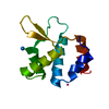
| ||||||||
|---|---|---|---|---|---|---|---|---|---|
| 1 |
| ||||||||
| Unit cell |
| ||||||||
| Details | The extent of the biological unit was not examined in this experiment. |
-Components
| #1: Protein | Mass: 13813.487 Da / Num. of mol.: 1 / Fragment: C-terminal domain (UNP residues 458-577) Source method: isolated from a genetically manipulated source Source: (gene. exp.) Homo sapiens (human) / Gene: ORC2, ORC2L / Plasmid: pET28-MHL / Production host:  | ||
|---|---|---|---|
| #2: Chemical |   Num. of mol.: 3 / Source method: obtained synthetically Num. of mol.: 3 / Source method: obtained synthetically#3: Water | ChemComp-HOH / |  Mass: 18.015 Da / Num. of mol.: 24 / Source method: isolated from a natural source / Formula: H2O Mass: 18.015 Da / Num. of mol.: 24 / Source method: isolated from a natural source / Formula: H2O |
-Experimental details
-Experiment
| Experiment | Method: X-RAY DIFFRACTION / Number of used crystals: 1 |
|---|
- Sample preparation
| Crystal | Density Matthews: 2.39 Å3/Da / Density % sol: 48.62 % |
|---|---|
| Crystal grow | Temperature: 293 K / Method: vapor diffusion, sitting drop / Details: 20% PEG-3350, 0.2 M magnesium formate |
-Data collection
| Diffraction | Mean temperature: 100 K | |||||||||||||||||||||||||||
|---|---|---|---|---|---|---|---|---|---|---|---|---|---|---|---|---|---|---|---|---|---|---|---|---|---|---|---|---|
| Diffraction source | Source: SYNCHROTRON / Site: APS  / Beamline: 19-ID / Wavelength: 0.9792604 Å / Beamline: 19-ID / Wavelength: 0.9792604 Å | |||||||||||||||||||||||||||
| Detector | Type: ADSC QUANTUM 315r / Detector: CCD / Date: Nov 28, 2014 | |||||||||||||||||||||||||||
| Radiation | Protocol: SINGLE WAVELENGTH / Monochromatic (M) / Laue (L): M / Scattering type: x-ray | |||||||||||||||||||||||||||
| Radiation wavelength | Wavelength: 0.9792604 Å / Relative weight: 1 | |||||||||||||||||||||||||||
| Reflection | Resolution: 2.01→47.66 Å / Num. obs: 8700 / % possible obs: 99.8 % / Redundancy: 11 % / CC1/2: 0.999 / Rmerge(I) obs: 0.077 / Rpim(I) all: 0.024 / Net I/σ(I): 21.5 / Num. measured all: 96076 | |||||||||||||||||||||||||||
| Reflection shell | Diffraction-ID: 1 / Rejects: _
|
-Phasing
| Phasing | Method: molecular replacement |
|---|
- Processing
| Software |
| |||||||||||||||||||||||||||||||||||||||||||||||||||||||||||||||||||||||||||
|---|---|---|---|---|---|---|---|---|---|---|---|---|---|---|---|---|---|---|---|---|---|---|---|---|---|---|---|---|---|---|---|---|---|---|---|---|---|---|---|---|---|---|---|---|---|---|---|---|---|---|---|---|---|---|---|---|---|---|---|---|---|---|---|---|---|---|---|---|---|---|---|---|---|---|---|---|
| Refinement | Method to determine structure: MOLECULAR REPLACEMENT Starting model: polyalanine coordinates derived from PDB entry 4XGC. Resolution: 2.01→47.66 Å / Cor.coef. Fo:Fc: 0.958 / Cor.coef. Fo:Fc free: 0.918 / WRfactor Rfree: 0.2429 / WRfactor Rwork: 0.1825 / FOM work R set: 0.8216 / SU B: 9.777 / SU ML: 0.132 / SU R Cruickshank DPI: 0.1792 / SU Rfree: 0.1801 / Cross valid method: THROUGHOUT / σ(F): 0 / ESU R: 0.179 / ESU R Free: 0.18 / Stereochemistry target values: MAXIMUM LIKELIHOOD Details: Data were reduced with HKL3000 for structure solution and early refinement. Molecular replacement was followed by simulated annealing refinement with PHENIX, subsequent phase improvement and ...Details: Data were reduced with HKL3000 for structure solution and early refinement. Molecular replacement was followed by simulated annealing refinement with PHENIX, subsequent phase improvement and automated model buidling with ARP/WARP. Diffraction data were reduced with XDS/AIMLESS for later refinement steps.
| |||||||||||||||||||||||||||||||||||||||||||||||||||||||||||||||||||||||||||
| Solvent computation | Ion probe radii: 0.8 Å / Shrinkage radii: 0.8 Å / VDW probe radii: 1.2 Å / Solvent model: MASK | |||||||||||||||||||||||||||||||||||||||||||||||||||||||||||||||||||||||||||
| Displacement parameters | Biso max: 92.98 Å2 / Biso mean: 39.091 Å2 / Biso min: 26.46 Å2
| |||||||||||||||||||||||||||||||||||||||||||||||||||||||||||||||||||||||||||
| Refinement step | Cycle: final / Resolution: 2.01→47.66 Å
| |||||||||||||||||||||||||||||||||||||||||||||||||||||||||||||||||||||||||||
| Refine LS restraints |
| |||||||||||||||||||||||||||||||||||||||||||||||||||||||||||||||||||||||||||
| LS refinement shell | Resolution: 2.012→2.064 Å / Total num. of bins used: 20
| |||||||||||||||||||||||||||||||||||||||||||||||||||||||||||||||||||||||||||
| Refinement TLS params. | Method: refined / Origin x: 35.851 Å / Origin y: 13.1035 Å / Origin z: -5.9789 Å
|
