 Movie
Movie Controller

[English] 日本語
 Yorodumi
- PDB-5bkd: Crystal structure of AAD-1 in complex with (R)-cyhalofop, Mn(II),... -
+ Open data

- Basic information
| Entry | Database: PDB / ID: 5bkd | ||||||
|---|---|---|---|---|---|---|---|
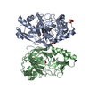
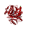
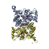
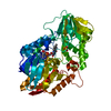
| Title | Crystal structure of AAD-1 in complex with (R)-cyhalofop, Mn(II), and 2-oxoglutarate | ||||||
 Components Components | (R)-phenoxypropionate/alpha-ketoglutarate-dioxygenase | ||||||
 Keywords Keywords | OXIDOREDUCTASE / dioxygenase / herbicide degradation | ||||||
| Function / homology |  Function and homology information Function and homology information(R)-dichlorprop dioxygenase (2-oxoglutarate) / 2-oxoglutarate-dependent dioxygenase activity / L-ascorbic acid binding / metal ion binding / cytoplasm Similarity search - Function | ||||||
| Biological species |  Delftia acidovorans (bacteria) Delftia acidovorans (bacteria) | ||||||
| Method |  X-RAY DIFFRACTION / SYNCHROTRON / MOLECULAR REPLACEMENT / Resolution: 1.9 Å X-RAY DIFFRACTION / SYNCHROTRON / MOLECULAR REPLACEMENT / Resolution: 1.9 Å | ||||||
 Authors Authors | Chekan, J.R. / Nair, S.K. | ||||||
 Citation Citation | Journal: Proc.Natl.Acad.Sci.USA / Year: 2019 Title: Molecular basis for enantioselective herbicide degradation imparted by aryloxyalkanoate dioxygenases in transgenic plants. Authors: Chekan, J.R. / Ongpipattanakul, C. / Wright, T.R. / Zhang, B. / Bollinger Jr., J.M. / Rajakovich, L.J. / Krebs, C. / Cicchillo, R.M. / Nair, S.K. | ||||||
| History |
|
- Structure visualization
| Structure viewer | Molecule: MolmilJmol/JSmol |
|---|
- Downloads & links
-Download
| PDBx/mmCIF format | 5bkd.cif.gz | 141.8 KB | Display | PDBx/mmCIF format |
|---|---|---|---|---|
| PDB format | pdb5bkd.ent.gz | 108 KB | Display | PDB format |
| PDBx/mmJSON format | 5bkd.json.gz | Tree view | PDBx/mmJSON format | |
| Others |  Other downloads Other downloads |
-Validation report
| Arichive directory | https://data.pdbj.org/pub/pdb/validation_reports/bk/5bkdftp://data.pdbj.org/pub/pdb/validation_reports/bk/5bkd | HTTPS FTP |
|---|
-Related structure data
| Related structure data | 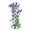 5bk9C  5bkbC  5bkcC  5bkeC  1gqwS C: citing same article ( S: Starting model for refinement |
|---|---|
| Similar structure data |


-Links
 PDBj
PDBj

- Assembly
| Deposited unit | 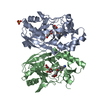
| ||||||||
|---|---|---|---|---|---|---|---|---|---|
| 1 |
| ||||||||
| Unit cell |
|
-Components
-Protein , 1 types, 2 molecules AB
| #1: Protein | Mass: 33252.578 Da / Num. of mol.: 2 Source method: isolated from a genetically manipulated source Source: (gene. exp.) Delftia acidovorans (bacteria) / Gene: rdpA / Production host: References: UniProt: P83310, (R)-dichlorprop dioxygenase (2-oxoglutarate) |
|---|
-Non-polymers , 5 types, 584 molecules 


| #2: Chemical |  Mass: 54.938 Da / Num. of mol.: 2 / Source method: obtained synthetically / Formula: Mn / Feature type: SUBJECT OF INVESTIGATION Mass: 54.938 Da / Num. of mol.: 2 / Source method: obtained synthetically / Formula: Mn / Feature type: SUBJECT OF INVESTIGATION#3: Chemical |  Mass: 146.098 Da / Num. of mol.: 2 / Source method: obtained synthetically / Formula: C5H6O5 / Feature type: SUBJECT OF INVESTIGATION Mass: 146.098 Da / Num. of mol.: 2 / Source method: obtained synthetically / Formula: C5H6O5 / Feature type: SUBJECT OF INVESTIGATION#4: Chemical |  Mass: 301.269 Da / Num. of mol.: 2 / Source method: obtained synthetically / Formula: C16H12FNO4 / Feature type: SUBJECT OF INVESTIGATION Mass: 301.269 Da / Num. of mol.: 2 / Source method: obtained synthetically / Formula: C16H12FNO4 / Feature type: SUBJECT OF INVESTIGATION#5: Chemical |  Mass: 96.063 Da / Num. of mol.: 2 / Source method: obtained synthetically / Formula: SO4 Mass: 96.063 Da / Num. of mol.: 2 / Source method: obtained synthetically / Formula: SO4#6: Water | ChemComp-HOH / | Mass: 18.015 Da / Num. of mol.: 576 / Source method: isolated from a natural source / Formula: H2O |
|---|
-Experimental details
-Experiment
| Experiment | Method: X-RAY DIFFRACTION / Number of used crystals: 1 |
|---|
- Sample preparation
| Crystal | Density Matthews: 2.19 Å3/Da / Density % sol: 43.74 % |
|---|---|
| Crystal grow | Temperature: 277 K / Method: vapor diffusion, hanging drop / pH: 9 Details: 25% PEG 3350 0.3 LiSO4 0.1M Bicine pH 9.0 8 mg/mL AAD-1 15% glycerol |
-Data collection
| Diffraction | Mean temperature: 100 K / Serial crystal experiment: N |
|---|---|
| Diffraction source | Source: SYNCHROTRON / Site: APS  / Beamline: 21-ID-F / Wavelength: 0.9787 Å / Beamline: 21-ID-F / Wavelength: 0.9787 Å |
| Detector | Type: MARMOSAIC 225 mm CCD / Detector: CCD / Date: Feb 23, 2014 |
| Radiation | Protocol: SINGLE WAVELENGTH / Monochromatic (M) / Laue (L): M / Scattering type: x-ray |
| Radiation wavelength | Wavelength: 0.9787 Å / Relative weight: 1 |
| Reflection | Resolution: 1.9→85.5 Å / Num. obs: 46644 / % possible obs: 100 % / Redundancy: 8.2 % / CC1/2: 0.998 / Rsym value: 0.119 / Net I/σ(I): 17 |
| Reflection shell | Resolution: 1.9→1.906 Å / Mean I/σ(I) obs: 2.9 / Num. unique obs: 460 / CC1/2: 0.81 / Rsym value: 0.755 / % possible all: 97.7 |
- Processing
| Software |
| |||||||||||||||||||||||||||||||||||||||||||||||||||||||||||||||||||||||||||||||||||||||||||||||||||||||||||||||||||||||
|---|---|---|---|---|---|---|---|---|---|---|---|---|---|---|---|---|---|---|---|---|---|---|---|---|---|---|---|---|---|---|---|---|---|---|---|---|---|---|---|---|---|---|---|---|---|---|---|---|---|---|---|---|---|---|---|---|---|---|---|---|---|---|---|---|---|---|---|---|---|---|---|---|---|---|---|---|---|---|---|---|---|---|---|---|---|---|---|---|---|---|---|---|---|---|---|---|---|---|---|---|---|---|---|---|---|---|---|---|---|---|---|---|---|---|---|---|---|---|---|---|
| Refinement | Method to determine structure: MOLECULAR REPLACEMENT Starting model: 1GQW Resolution: 1.9→56.742 Å / SU ML: 0.19 / Cross valid method: FREE R-VALUE / σ(F): 1.34 / Phase error: 20.78
| |||||||||||||||||||||||||||||||||||||||||||||||||||||||||||||||||||||||||||||||||||||||||||||||||||||||||||||||||||||||
| Solvent computation | Shrinkage radii: 0.9 Å / VDW probe radii: 1.11 Å | |||||||||||||||||||||||||||||||||||||||||||||||||||||||||||||||||||||||||||||||||||||||||||||||||||||||||||||||||||||||
| Refinement step | Cycle: LAST / Resolution: 1.9→56.742 Å
| |||||||||||||||||||||||||||||||||||||||||||||||||||||||||||||||||||||||||||||||||||||||||||||||||||||||||||||||||||||||
| Refine LS restraints |
| |||||||||||||||||||||||||||||||||||||||||||||||||||||||||||||||||||||||||||||||||||||||||||||||||||||||||||||||||||||||
| LS refinement shell |
|
