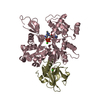
 Movie
Movie Controller


+ Open data

- Basic information
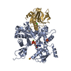
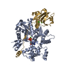
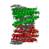
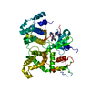
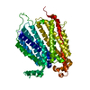
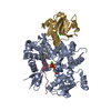
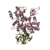
| Entry | Database: PDB / ID: 5b5r | ||||||
|---|---|---|---|---|---|---|---|
| Title | Crystal structure of GSDMA3 | ||||||
 Components Components | Gasdermin-A3 | ||||||
 Keywords Keywords |  TRANSPORT PROTEIN / pyroptosis excutioner TRANSPORT PROTEIN / pyroptosis excutioner | ||||||
| Function / homology |  Function and homology information Function and homology informationsebaceous gland cell differentiation / avascular cornea development in camera-type eye / negative regulation of timing of anagen / hair cycle / wide pore channel activity / mammary gland development / phosphatidylinositol-4-phosphate binding / positive regulation of extrinsic apoptotic signaling pathway / skin development / hair follicle morphogenesis ...sebaceous gland cell differentiation / avascular cornea development in camera-type eye / negative regulation of timing of anagen / hair cycle / wide pore channel activity / mammary gland development / phosphatidylinositol-4-phosphate binding / positive regulation of extrinsic apoptotic signaling pathway / skin development / hair follicle morphogenesis / phosphatidylserine binding / pyroptosis / somatic stem cell population maintenance / hair follicle development / extrinsic apoptotic signaling pathway / phosphatidylinositol-4,5-bisphosphate binding / positive regulation of interleukin-1 beta production / mitochondrial membrane / negative regulation of canonical Wnt signaling pathway / defense response to bacterium / positive regulation of apoptotic process / perinuclear region of cytoplasm / membrane / nucleus / plasma membrane / cytosolSimilarity search - Function | ||||||
| Biological species |  Mus musculus (house mouse) Mus musculus (house mouse) | ||||||
| Method | X-RAY DIFFRACTION / SYNCHROTRON / SAD / Resolution: 1.902 Å | ||||||
 Authors Authors | Ding, J. / Shao, F. | ||||||
 Citation Citation | Journal: Nature / Year: 2016 Title: Pore-forming activity and structural autoinhibition of the gasdermin family. Authors: Ding, J. / Wang, K. / Liu, W. / She, Y. / Sun, Q. / Shi, J. / Sun, H. / Wang, D.C. / Shao, F. | ||||||
| History |
|
- Structure visualization
| Structure viewer | Molecule: MolmilJmol/JSmol |
|---|
- Downloads & links
-Download
| PDBx/mmCIF format | 5b5r.cif.gz | 97.9 KB | Display | PDBx/mmCIF format |
|---|---|---|---|---|
| PDB format | pdb5b5r.ent.gz | 76.7 KB | Display | PDB format |
| PDBx/mmJSON format | 5b5r.json.gz | Tree view | PDBx/mmJSON format | |
| Others |  Other downloads Other downloads |
-Validation report
| Arichive directory | https://data.pdbj.org/pub/pdb/validation_reports/b5/5b5rftp://data.pdbj.org/pub/pdb/validation_reports/b5/5b5r | HTTPS FTP |
|---|
-Related structure data
| Similar structure data |
|---|
-Links
 PDBj
PDBj- Assembly
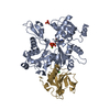
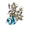
| Deposited unit | 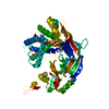
| ||||||||
|---|---|---|---|---|---|---|---|---|---|
| 1 |
| ||||||||
| Unit cell |
|
-Components
| #1: Protein | Mass: 52854.457 Da / Num. of mol.: 1 Source method: isolated from a genetically manipulated source Source: (gene. exp.) Mus musculus (house mouse) / Gene: Gsdma3, Gsdm3 / Plasmid: pSUMO / Production host:  Escherichia coli (E. coli) / Strain (production host): BL21(DE3) / References: UniProt: Q5Y4Y6 Escherichia coli (E. coli) / Strain (production host): BL21(DE3) / References: UniProt: Q5Y4Y6 |
|---|---|
| #2: Water | ChemComp-HOH / Water Mass: 18.015 Da / Num. of mol.: 218 / Source method: isolated from a natural source / Formula: H2O Mass: 18.015 Da / Num. of mol.: 218 / Source method: isolated from a natural source / Formula: H2O |
-Experimental details
-Experiment
| Experiment | Method: X-RAY DIFFRACTION / Number of used crystals: 1 |
|---|
- Sample preparation
| Crystal | Density Matthews: 1.99 Å3/Da / Density % sol: 38.16 % Description: THE ENTRY CONTAINS FRIEDEL PAIRS IN I_PLUS/MINUS COLUMNS |
|---|---|
| Crystal grow | Temperature: 293 K / Method: vapor diffusion, sitting drop / pH: 6.5 Details: 100 mM Bis-Tris (pH 6.5), 19% polyethylene glycol 3550, 10 mM TCEP |
-Data collection
| Diffraction | Mean temperature: 95 K |
|---|---|
| Diffraction source | Source: SYNCHROTRON / Site: SSRF  / Beamline: BL19U1 / Wavelength: 0.97776 Å / Beamline: BL19U1 / Wavelength: 0.97776 Å |
| Detector | Type: DECTRIS PILATUS3 6M / Detector: PIXEL / Date: Sep 6, 2015 |
| Radiation | Protocol: SINGLE WAVELENGTH / Monochromatic (M) / Laue (L): M / Scattering type: x-ray |
| Radiation wavelength | Wavelength: 0.97776 Å / Relative weight: 1 |
| Reflection | Resolution: 1.9→50 Å / Num. obs: 44110 / % possible obs: 97.1 % / Redundancy: 6.6 % / Rmerge(I) obs: 0.071 / Net I/σ(I): 25.6 |
| Reflection shell | Resolution: 1.9→1.93 Å / Redundancy: 5.1 % / Rmerge(I) obs: 0.876 / Mean I/σ(I) obs: 2.4 / % possible all: 92.7 |
- Processing
| Software |
| |||||||||||||||||||||||||||||||||||||||||||||||||||||||||||||||||||||||||||||||||||||||||||||||||||||||||||||||||||||||
|---|---|---|---|---|---|---|---|---|---|---|---|---|---|---|---|---|---|---|---|---|---|---|---|---|---|---|---|---|---|---|---|---|---|---|---|---|---|---|---|---|---|---|---|---|---|---|---|---|---|---|---|---|---|---|---|---|---|---|---|---|---|---|---|---|---|---|---|---|---|---|---|---|---|---|---|---|---|---|---|---|---|---|---|---|---|---|---|---|---|---|---|---|---|---|---|---|---|---|---|---|---|---|---|---|---|---|---|---|---|---|---|---|---|---|---|---|---|---|---|---|
| Refinement | Method to determine structure: SAD / Resolution: 1.902→37.914 Å / SU ML: 0.21 / Cross valid method: FREE R-VALUE / σ(F): 1.38 / Phase error: 25.31 Details: THE ENTRY CONTAINS FRIEDEL PAIRS IN I_PLUS/MINUS COLUMNS
| |||||||||||||||||||||||||||||||||||||||||||||||||||||||||||||||||||||||||||||||||||||||||||||||||||||||||||||||||||||||
| Solvent computation | Shrinkage radii: 0.9 Å / VDW probe radii: 1.11 Å | |||||||||||||||||||||||||||||||||||||||||||||||||||||||||||||||||||||||||||||||||||||||||||||||||||||||||||||||||||||||
| Refinement step | Cycle: LAST / Resolution: 1.902→37.914 Å
| |||||||||||||||||||||||||||||||||||||||||||||||||||||||||||||||||||||||||||||||||||||||||||||||||||||||||||||||||||||||
| Refine LS restraints |
| |||||||||||||||||||||||||||||||||||||||||||||||||||||||||||||||||||||||||||||||||||||||||||||||||||||||||||||||||||||||
| LS refinement shell |
|
