 Movie
Movie Controller

[English] 日本語
 Yorodumi
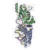
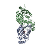
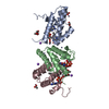
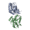
- PDB-5a77: Crystal structure of the homing endonuclease I-CvuI in complex wi... -
+ Open data

- Basic information
| Entry | Database: PDB / ID: 5a77 | ||||||
|---|---|---|---|---|---|---|---|
| Title | Crystal structure of the homing endonuclease I-CvuI in complex with I- CreI target (C1221) in the presence of 2 mM Mg revealing DNA cleaved | ||||||
 Components Components |
| ||||||
 Keywords Keywords | HYDROLASE/DNA / HYDROLASE-DNA COMPLEX / GENE TARGETING / GENETICS / PROTEIN-DNA INTERACTION / HOMING ENDONUCLEASES | ||||||
| Function / homology |  Function and homology information Function and homology informationintron homing / chloroplast / endonuclease activity / Hydrolases; Acting on ester bonds Similarity search - Function | ||||||
| Biological species |  CHLORELLA VULGARIS (plant) CHLORELLA VULGARIS (plant)SYNTHETIC CONSTRUCT (others) | ||||||
| Method |  X-RAY DIFFRACTION / SYNCHROTRON / MOLECULAR REPLACEMENT / Resolution: 2.5 Å X-RAY DIFFRACTION / SYNCHROTRON / MOLECULAR REPLACEMENT / Resolution: 2.5 Å | ||||||
 Authors Authors | Molina, R. / Redondo, P. / LopezMendez, B. / Villate, M. / Merino, N. / Blanco, F.J. / Valton, J. / Grizot, S. / Duchateau, P. / Prieto, J. / Montoya, G. | ||||||
 Citation Citation | Journal: J.Biol.Chem. / Year: 2015 Title: Crystal Structure of the Homing Endonuclease I-Cvui Provides a New Template for Genome Modification Authors: Molina, R. / Redondo, P. / Lopez-Mendez, B. / Villate, M. / Merino, N. / Blanco, F.J. / Valton, J. / Grizot, S. / Duchateau, P. / Prieto, J. / Montoya, G. | ||||||
| History |
|
- Structure visualization
| Structure viewer | Molecule: MolmilJmol/JSmol |
|---|
- Downloads & links
-Download
| PDBx/mmCIF format | 5a77.cif.gz | 102.6 KB | Display | PDBx/mmCIF format |
|---|---|---|---|---|
| PDB format | pdb5a77.ent.gz | 74.5 KB | Display | PDB format |
| PDBx/mmJSON format | 5a77.json.gz | Tree view | PDBx/mmJSON format | |
| Others |  Other downloads Other downloads |
-Validation report
| Summary document | 5a77_validation.pdf.gz | 448.4 KB | Display | wwPDB validaton report |
|---|---|---|---|---|
| Full document | 5a77_full_validation.pdf.gz | 454 KB | Display | |
| Data in XML | 5a77_validation.xml.gz | 14.8 KB | Display | |
| Data in CIF | 5a77_validation.cif.gz | 19.9 KB | Display | |
| Arichive directory | https://data.pdbj.org/pub/pdb/validation_reports/a7/5a77ftp://data.pdbj.org/pub/pdb/validation_reports/a7/5a77 | HTTPS FTP |
-Related structure data
| Related structure data | 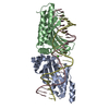 5a72C  5a74C 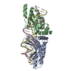 5a78C  1g9zS  5a73 5a75 C: citing same article ( S: Starting model for refinement |
|---|---|
| Similar structure data |


-Links
 PDBj
PDBj


- Assembly
| Deposited unit | 
| ||||||||
|---|---|---|---|---|---|---|---|---|---|
| 1 |
| ||||||||
| Unit cell |
|
-Components
| #1: Protein | Mass: 19292.533 Da / Num. of mol.: 2 Source method: isolated from a genetically manipulated source Source: (gene. exp.) CHLORELLA VULGARIS (plant) / Production host:  References: UniProt: P56347, Hydrolases; Acting on ester bonds #2: DNA chain | Mass: 4248.793 Da / Num. of mol.: 2 / Source method: obtained synthetically / Source: (synth.) SYNTHETIC CONSTRUCT (others) #3: DNA chain | Mass: 3075.026 Da / Num. of mol.: 2 / Source method: obtained synthetically / Source: (synth.) SYNTHETIC CONSTRUCT (others) #4: Chemical |   Mass: 24.305 Da / Num. of mol.: 2 / Source method: obtained synthetically / Formula: Mg Mass: 24.305 Da / Num. of mol.: 2 / Source method: obtained synthetically / Formula: Mg#5: Water | ChemComp-HOH / |  Mass: 18.015 Da / Num. of mol.: 17 / Source method: isolated from a natural source / Formula: H2O Mass: 18.015 Da / Num. of mol.: 17 / Source method: isolated from a natural source / Formula: H2O |
|---|
-Experimental details
-Experiment
| Experiment | Method: X-RAY DIFFRACTION / Number of used crystals: 1 |
|---|
- Sample preparation
| Crystal | Density Matthews: 2.34 Å3/Da / Density % sol: 53 % / Description: NONE |
|---|
-Data collection
| Diffraction | Mean temperature: 100 K |
|---|---|
| Diffraction source | Source: SYNCHROTRON / Site: SLS  / Beamline: X06SA / Wavelength: 1 / Beamline: X06SA / Wavelength: 1 |
| Detector | Type: DECTRIS PILATUS 2M / Detector: PIXEL |
| Radiation | Protocol: SINGLE WAVELENGTH / Monochromatic (M) / Laue (L): M / Scattering type: x-ray |
| Radiation wavelength | Wavelength: 1 Å / Relative weight: 1 |
| Reflection | Resolution: 2.5→47.54 Å / Num. obs: 15872 / % possible obs: 99.2 % / Observed criterion σ(I): 2 / Redundancy: 4.8 % / Biso Wilson estimate: 36.8 Å2 / Rmerge(I) obs: 0.12 / Net I/σ(I): 7.1 |
| Reflection shell | Resolution: 2.5→2.64 Å / Redundancy: 4.7 % / Rmerge(I) obs: 0.41 / Mean I/σ(I) obs: 2.7 / % possible all: 99 |
- Processing
| Software |
| |||||||||||||||||||||||||||||||||||||||||||||||||||||||||||||||||||||||||||||||||||||||||||||||||||||||||||||||||||||||||||||||||||||||||||||||||||||||||||||||||
|---|---|---|---|---|---|---|---|---|---|---|---|---|---|---|---|---|---|---|---|---|---|---|---|---|---|---|---|---|---|---|---|---|---|---|---|---|---|---|---|---|---|---|---|---|---|---|---|---|---|---|---|---|---|---|---|---|---|---|---|---|---|---|---|---|---|---|---|---|---|---|---|---|---|---|---|---|---|---|---|---|---|---|---|---|---|---|---|---|---|---|---|---|---|---|---|---|---|---|---|---|---|---|---|---|---|---|---|---|---|---|---|---|---|---|---|---|---|---|---|---|---|---|---|---|---|---|---|---|---|---|---|---|---|---|---|---|---|---|---|---|---|---|---|---|---|---|---|---|---|---|---|---|---|---|---|---|---|---|---|---|---|---|
| Refinement | Method to determine structure: MOLECULAR REPLACEMENT Starting model: PDB ENTRY 1G9Z Resolution: 2.5→47.539 Å / SU ML: 0.3 / σ(F): 1.28 / Phase error: 37 / Stereochemistry target values: ML
| |||||||||||||||||||||||||||||||||||||||||||||||||||||||||||||||||||||||||||||||||||||||||||||||||||||||||||||||||||||||||||||||||||||||||||||||||||||||||||||||||
| Solvent computation | Shrinkage radii: 0.9 Å / VDW probe radii: 1.11 Å / Solvent model: FLAT BULK SOLVENT MODEL | |||||||||||||||||||||||||||||||||||||||||||||||||||||||||||||||||||||||||||||||||||||||||||||||||||||||||||||||||||||||||||||||||||||||||||||||||||||||||||||||||
| Refinement step | Cycle: LAST / Resolution: 2.5→47.539 Å
| |||||||||||||||||||||||||||||||||||||||||||||||||||||||||||||||||||||||||||||||||||||||||||||||||||||||||||||||||||||||||||||||||||||||||||||||||||||||||||||||||
| Refine LS restraints |
| |||||||||||||||||||||||||||||||||||||||||||||||||||||||||||||||||||||||||||||||||||||||||||||||||||||||||||||||||||||||||||||||||||||||||||||||||||||||||||||||||
| LS refinement shell |
|
