 Movie
Movie Controller


+ Open data

- Basic information
| Entry | Database: PDB / ID: 4xqw | |||||||||
|---|---|---|---|---|---|---|---|---|---|---|
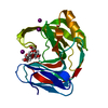
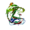
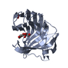
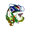
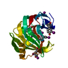
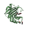
| Title | X-ray structure analysis of xylanase-N44E with MES at pH6.0 | |||||||||
 Components Components | Endo-1,4-beta-xylanase 2 Xylanase Xylanase | |||||||||
 Keywords Keywords | HYDROLASE / jelly roll | |||||||||
| Function / homology |  Function and homology informationendo-1,4-beta-xylanase activity / endo-1,4-beta-xylanase / xylan catabolic process / extracellular region Function and homology informationendo-1,4-beta-xylanase activity / endo-1,4-beta-xylanase / xylan catabolic process / extracellular regionSimilarity search - Function | |||||||||
| Biological species |  Hypocrea jecorina (fungus) Hypocrea jecorina (fungus) | |||||||||
| Method | X-RAY DIFFRACTION / MOLECULAR REPLACEMENT / Resolution: 1.5 Å | |||||||||
 Authors Authors | Wan, Q. / Park, J.M. / Riccardi, D.M. / Hanson, L.B. / Fisher, Z. / Smith, J.C. / Ostermann, A. / Schrader, T. / Graham, D.E. / Coates, L. ...Wan, Q. / Park, J.M. / Riccardi, D.M. / Hanson, L.B. / Fisher, Z. / Smith, J.C. / Ostermann, A. / Schrader, T. / Graham, D.E. / Coates, L. / Langan, P. / Kovalevsky, A.Y. | |||||||||
| Funding support |  United States, United States,  China, 2items China, 2items
| |||||||||
 Citation Citation | Journal: Proc.Natl.Acad.Sci.USA / Year: 2015 Title: Direct determination of protonation states and visualization of hydrogen bonding in a glycoside hydrolase with neutron crystallography. Authors: Wan, Q. / Parks, J.M. / Hanson, B.L. / Fisher, S.Z. / Ostermann, A. / Schrader, T.E. / Graham, D.E. / Coates, L. / Langan, P. / Kovalevsky, A. #1: Journal: Acta Crystallogr.,Sect.F / Year: 2011 Title: Preliminary joint X-ray and neutron protein crystallographic studies of endoxylanase II from the fungus Trichoderma longibrachiatum. Authors: Kovalevsky, A.Y. / Hanson, B.L. / Seaver, S. / Fisher, S.Z. / Mustyakimov, M. / Langan, P. #2: Journal: Acta Crystallogr.,Sect.D / Year: 2014Title: X-ray crystallographic studies of family 11 xylanase Michaelis and product complexes: implications for the catalytic mechanism. Authors: Wan, Q. / Zhang, Q. / Hamilton-Brehm, S. / Weiss, K. / Mustyakimov, M. / Coates, L. / Langan, P. / Graham, D. / Kovalevsky, A. #3: Journal: Acta Crystallogr.,Sect.F / Year: 2013 Title: Heterologous expression, purification, crystallization and preliminary X-ray analysis of Trichoderma reesei xylanase II and four variants. Authors: Wan, Q. / Kovalevsky, A. / Zhang, Q. / Hamilton-Brehm, S. / Upton, R. / Weiss, K.L. / Mustyakimov, M. / Graham, D. / Coates, L. / Langan, P. | |||||||||
| History |
|
- Structure visualization
| Structure viewer | Molecule: MolmilJmol/JSmol |
|---|
- Downloads & links
-Download
| PDBx/mmCIF format | 4xqw.cif.gz | 58.4 KB | Display | PDBx/mmCIF format |
|---|---|---|---|---|
| PDB format | pdb4xqw.ent.gz | 39.4 KB | Display | PDB format |
| PDBx/mmJSON format | 4xqw.json.gz | Tree view | PDBx/mmJSON format | |
| Others |  Other downloads Other downloads |
-Validation report
| Arichive directory | https://data.pdbj.org/pub/pdb/validation_reports/xq/4xqwftp://data.pdbj.org/pub/pdb/validation_reports/xq/4xqw | HTTPS FTP |
|---|
-Related structure data
| Related structure data | 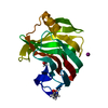 4s2dC 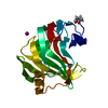 4s2fC 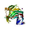 4s2gC 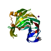 4s2hC 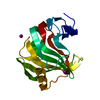 4xpvC 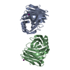 4xq4C 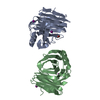 4xqdC  2dfbS C: citing same article ( S: Starting model for refinement |
|---|---|
| Similar structure data |


-Links
 PDBj
PDBj
- Assembly
| Deposited unit | 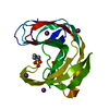
| ||||||||
|---|---|---|---|---|---|---|---|---|---|
| 1 |
| ||||||||
| Unit cell |
| ||||||||
| Details | biological unit is the same as asym. |
-Components
| #1: Protein | Xylanase / Xylanase 2 / 1 / 4-beta-D-xylan xylanohydrolase 2 Mass: 20742.348 Da / Num. of mol.: 1 / Fragment: substrate-binding groove, jelly roll Source method: isolated from a genetically manipulated source Source: (gene. exp.) Hypocrea jecorina (fungus) / Gene: xyn2 / Plasmid: pJexpress401 / Production host:  Escherichia coli (E. coli) / Strain (production host): BL21-Gold / References: UniProt: P36217, endo-1,4-beta-xylanase Escherichia coli (E. coli) / Strain (production host): BL21-Gold / References: UniProt: P36217, endo-1,4-beta-xylanase | ||||
|---|---|---|---|---|---|
| #2: Chemical | ChemComp-IOD / Iodide  Mass: 126.904 Da / Num. of mol.: 5 / Source method: obtained synthetically / Formula: I Mass: 126.904 Da / Num. of mol.: 5 / Source method: obtained synthetically / Formula: I#3: Chemical | ChemComp-MES / | MES (buffer)  Mass: 195.237 Da / Num. of mol.: 1 / Source method: obtained synthetically / Formula: C6H13NO4S / Comment: pH buffer*YM Mass: 195.237 Da / Num. of mol.: 1 / Source method: obtained synthetically / Formula: C6H13NO4S / Comment: pH buffer*YM#4: Water | ChemComp-HOH / | Water Mass: 18.015 Da / Num. of mol.: 234 / Source method: isolated from a natural source / Formula: H2O Mass: 18.015 Da / Num. of mol.: 234 / Source method: isolated from a natural source / Formula: H2O |
-Experimental details
-Experiment
| Experiment | Method: X-RAY DIFFRACTION / Number of used crystals: 1 |
|---|
- Sample preparation
| Crystal | Density Matthews: 2.4 Å3/Da / Density % sol: 48.72 % |
|---|---|
| Crystal grow | Temperature: 291 K / Method: vapor diffusion, hanging drop / pH: 6 / Details: 16% PEG8000, 0.2 M NaI, 0.1 M MES-NaOH at pH 6.0 |
-Data collection
| Diffraction | Mean temperature: 100 K |
|---|---|
| Diffraction source | Source: ROTATING ANODE / Type: RIGAKU MICROMAX-007 HF / Wavelength: 1.54 Å |
| Detector | Type: RIGAKU RAXIS IV++ / Detector: IMAGE PLATE / Date: Apr 12, 2013 / Details: Mirrors |
| Radiation | Monochromator: Ni Filter / Protocol: SINGLE WAVELENGTH / Monochromatic (M) / Laue (L): M / Scattering type: x-ray |
| Radiation wavelength | Wavelength: 1.54 Å / Relative weight: 1 |
| Reflection | Resolution: 1.5→50 Å / Num. obs: 31754 / % possible obs: 96.5 % / Redundancy: 3.8 % / Rsym value: 0.066 / Net I/σ(I): 17 |
| Reflection shell | Resolution: 1.5→1.53 Å / Redundancy: 3.8 % / Rmerge(I) obs: 0.427 / Mean I/σ(I) obs: 3.1 / % possible all: 92.7 |
- Processing
| Software |
| ||||||||||||||||||||||||||||||||||||||||||||||||||||||||||||||||||||||||||||||||||||
|---|---|---|---|---|---|---|---|---|---|---|---|---|---|---|---|---|---|---|---|---|---|---|---|---|---|---|---|---|---|---|---|---|---|---|---|---|---|---|---|---|---|---|---|---|---|---|---|---|---|---|---|---|---|---|---|---|---|---|---|---|---|---|---|---|---|---|---|---|---|---|---|---|---|---|---|---|---|---|---|---|---|---|---|---|---|
| Refinement | Method to determine structure: MOLECULAR REPLACEMENT Starting model: 2DFB Resolution: 1.5→32.93 Å / FOM work R set: 0.8898 / SU ML: 0.17 / Cross valid method: FREE R-VALUE / σ(F): 1.34 / Phase error: 17.63 / Stereochemistry target values: ML
| ||||||||||||||||||||||||||||||||||||||||||||||||||||||||||||||||||||||||||||||||||||
| Solvent computation | Shrinkage radii: 0.9 Å / VDW probe radii: 1.11 Å / Solvent model: FLAT BULK SOLVENT MODEL | ||||||||||||||||||||||||||||||||||||||||||||||||||||||||||||||||||||||||||||||||||||
| Displacement parameters | Biso max: 41.02 Å2 / Biso mean: 19.5 Å2 / Biso min: 11.76 Å2 | ||||||||||||||||||||||||||||||||||||||||||||||||||||||||||||||||||||||||||||||||||||
| Refinement step | Cycle: final / Resolution: 1.5→32.93 Å
| ||||||||||||||||||||||||||||||||||||||||||||||||||||||||||||||||||||||||||||||||||||
| Refine LS restraints |
| ||||||||||||||||||||||||||||||||||||||||||||||||||||||||||||||||||||||||||||||||||||
| LS refinement shell |
|
