 Movie
Movie Controller


+ Open data

- Basic information
| Entry | Database: PDB / ID: 4xpv | |||||||||
|---|---|---|---|---|---|---|---|---|---|---|
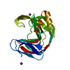
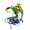
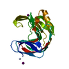
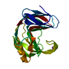
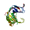
| Title | Neutron and X-ray structure analysis of xylanase: N44D at pH6 | |||||||||
 Components Components | Endo-1,4-beta-xylanase 2 | |||||||||
 Keywords Keywords | HYDROLASE / jelly roll | |||||||||
| Function / homology |  Function and homology information Function and homology informationendo-1,4-beta-xylanase activity / endo-1,4-beta-xylanase / xylan catabolic process / extracellular region Similarity search - Function | |||||||||
| Biological species |  Hypocrea jecorina (fungus) Hypocrea jecorina (fungus) | |||||||||
| Method |  X-RAY DIFFRACTION / NEUTRON DIFFRACTION / NUCLEAR REACTOR / MOLECULAR REPLACEMENT / Resolution: 1.7 Å X-RAY DIFFRACTION / NEUTRON DIFFRACTION / NUCLEAR REACTOR / MOLECULAR REPLACEMENT / Resolution: 1.7 Å | |||||||||
 Authors Authors | Wan, Q. / Park, J.M. / Riccardi, D.M. / Hanson, L.B. / Fisher, Z. / Smith, J.C. / Ostermann, A. / Schrader, T. / Graham, D.E. / Coates, L. ...Wan, Q. / Park, J.M. / Riccardi, D.M. / Hanson, L.B. / Fisher, Z. / Smith, J.C. / Ostermann, A. / Schrader, T. / Graham, D.E. / Coates, L. / Langan, P. / Kovalevsky, A.Y. | |||||||||
| Funding support |  United States, United States,  China, 2items China, 2items
| |||||||||
 Citation Citation | Journal: Proc.Natl.Acad.Sci.USA / Year: 2015 Title: Direct determination of protonation states and visualization of hydrogen bonding in a glycoside hydrolase with neutron crystallography. Authors: Wan, Q. / Parks, J.M. / Hanson, B.L. / Fisher, S.Z. / Ostermann, A. / Schrader, T.E. / Graham, D.E. / Coates, L. / Langan, P. / Kovalevsky, A. #1: Journal: Acta Crystallogr.,Sect.F / Year: 2011 Title: Preliminary joint X-ray and neutron protein crystallographic studies of endoxylanase II from the fungus Trichoderma longibrachiatum. Authors: Kovalevsky, A.Y. / Hanson, B.L. / Seaver, S. / Fisher, S.Z. / Mustyakimov, M. / Langan, P. #2: Journal: Acta Crystallogr.,Sect.D / Year: 2014Title: X-ray crystallographic studies of family 11 xylanase Michaelis and product complexes: implications for the catalytic mechanism. Authors: Wan, Q. / Zhang, Q. / Hamilton-Brehm, S. / Weiss, K. / Mustyakimov, M. / Coates, L. / Langan, P. / Graham, D. / Kovalevsky, A. #3: Journal: Acta Crystallogr.,Sect.F / Year: 2013 Title: Heterologous expression, purification, crystallization and preliminary X-ray analysis of Trichoderma reesei xylanase II and four variants. Authors: Wan, Q. / Kovalevsky, A. / Zhang, Q. / Hamilton-Brehm, S. / Upton, R. / Weiss, K.L. / Mustyakimov, M. / Graham, D. / Coates, L. / Langan, P. | |||||||||
| History |
|
- Structure visualization
| Structure viewer | Molecule: MolmilJmol/JSmol |
|---|
- Downloads & links
-Download
| PDBx/mmCIF format | 4xpv.cif.gz | 96.5 KB | Display | PDBx/mmCIF format |
|---|---|---|---|---|
| PDB format | pdb4xpv.ent.gz | 75.2 KB | Display | PDB format |
| PDBx/mmJSON format | 4xpv.json.gz | Tree view | PDBx/mmJSON format | |
| Others |  Other downloads Other downloads |
-Validation report
| Arichive directory | https://data.pdbj.org/pub/pdb/validation_reports/xp/4xpvftp://data.pdbj.org/pub/pdb/validation_reports/xp/4xpv | HTTPS FTP |
|---|
-Related structure data
| Related structure data | 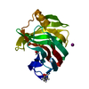 4s2dC 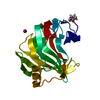 4s2fC 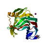 4s2gC 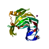 4s2hC 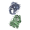 4xq4C 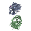 4xqdC  4xqwC 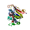 1rx2S S: Starting model for refinement C: citing same article ( |
|---|---|
| Similar structure data |


-Links
 PDBj
PDBj


- Assembly
| Deposited unit | 
| ||||||||
|---|---|---|---|---|---|---|---|---|---|
| 1 |
| ||||||||
| Unit cell |
|
-Components
| #1: Protein | Mass: 20728.322 Da / Num. of mol.: 1 / Mutation: N44D Source method: isolated from a genetically manipulated source Source: (gene. exp.) Hypocrea jecorina (fungus) / Gene: xyn2 / Plasmid: pJexpress401 / Production host:  | ||||
|---|---|---|---|---|---|
| #2: Chemical |   Mass: 126.904 Da / Num. of mol.: 3 / Source method: obtained synthetically / Formula: I Mass: 126.904 Da / Num. of mol.: 3 / Source method: obtained synthetically / Formula: I#3: Chemical | ChemComp-DOD / |   Mass: 18.015 Da / Num. of mol.: 173 / Source method: isolated from a natural source / Formula: D2O Mass: 18.015 Da / Num. of mol.: 173 / Source method: isolated from a natural source / Formula: D2OHas protein modification | N | |
-Experimental details
-Experiment
| Experiment |
|
|---|
- Sample preparation
| Crystal | Density Matthews: 2.52 Å3/Da / Density % sol: 51.24 % |
|---|---|
| Crystal grow | Temperature: 291 K / Method: vapor diffusion, sitting drop / pH: 7 / Details: 16% PEG8000, 0.2 M NaI, 0.1 M HEPES at pH 7.0 / PH range: 6-7 |
-Data collection
| Diffraction |
| |||||||||||||||||||||
|---|---|---|---|---|---|---|---|---|---|---|---|---|---|---|---|---|---|---|---|---|---|---|
| Diffraction source |
| |||||||||||||||||||||
| Detector |
| |||||||||||||||||||||
| Radiation |
| |||||||||||||||||||||
| Radiation wavelength |
| |||||||||||||||||||||
| Reflection | Entry-ID: 4XPV
| |||||||||||||||||||||
| Reflection shell |
|
- Processing
| Software |
| |||||||||||||||||||||||||||||||||
|---|---|---|---|---|---|---|---|---|---|---|---|---|---|---|---|---|---|---|---|---|---|---|---|---|---|---|---|---|---|---|---|---|---|---|
| Refinement | % reflection Rfree: 5 % / R Free selection details: RANDOM / Cross valid method: FREE R-VALUE
| |||||||||||||||||||||||||||||||||
| Refinement step | Cycle: final / Resolution: 1.7→20 Å
| |||||||||||||||||||||||||||||||||
| Refine LS restraints |
| |||||||||||||||||||||||||||||||||
| LS refinement shell |
|
