[English] 日本語
Yorodumi
- PDB-4uid: Crystal structure of the S-layer protein SbsC domains 4 and 5 -

+
Open data


ID or keywords:

Loading...

-
Basic information

Entry
Database: PDB / ID: 4uid
TitleCrystal structure of the S-layer protein SbsC domains 4 and 5
ComponentsSURFACE LAYER PROTEIN
KeywordsSUGAR BINDING PROTEIN / S-LAYER / SBSC / G. STEAROTHERMOPHILUS
Function / homology
Function and homology information


identical protein binding / metal ion binding
Similarity search - Function
S-layer protein SbsC, C-terminal domain / SbsC C-terminal domain / SbsC C-terminal domain / S-layer protein SbsC C-terminal domain / Copper resistance protein CopC/internalin, immunoglobulin-like / Bacterial Ig-like domain 2 / Bacterial Ig-like domain, group 2 / Immunoglobulin-like fold
Similarity search - Domain/homology
Surface layer protein
Similarity search - Component
Biological speciesGEOBACILLUS STEAROTHERMOPHILUS (bacteria)
MethodX-RAY DIFFRACTION / SYNCHROTRON / MOLECULAR REPLACEMENT / Resolution: 2.8 Å
AuthorsPavkov-Keller, T. / Dordic, A. / Egelseer, E.M. / Sleytr, U.B. / Keller, W.
CitationJournal: To be Published
Title: Structure of the S-Layer Protein Sbsc
Authors: Pavkov-Keller, T. / Dordic, A. / Eder, M. / Davies, K. / Mills, D. / Egelseer, E.M. / Sleytr, U.B. / Kuehlbrandt, W. / Vonck, J. / Keller, W.
History
DepositionMar 27, 2015Deposition site: PDBE / Processing site: PDBE
Revision 1.0Apr 13, 2016Provider: repository / Type: Initial release

-
Structure visualization

Structure viewerMolecule:
MolmilJmol/JSmol

Downloads & links

-
Assembly

Deposited unit
A: SURFACE LAYER PROTEIN
B: SURFACE LAYER PROTEIN


Theoretical massNumber of molelcules
Total (without water)43,2842
Polymers43,2842
Non-polymers00
Water1,06359
1
A: SURFACE LAYER PROTEIN


Theoretical massNumber of molelcules
Total (without water)21,6421
Polymers21,6421
Non-polymers00
Water181
TypeNameSymmetry operationNumber
identity operation1_555x,y,z1
MethodPISA
2
B: SURFACE LAYER PROTEIN


Theoretical massNumber of molelcules
Total (without water)21,6421
Polymers21,6421
Non-polymers00
Water181
TypeNameSymmetry operationNumber
identity operation1_555x,y,z1
MethodPISA
Unit cell
Length a, b, c (Å)110.367, 110.367, 87.143
Angle α, β, γ (deg.)90.00, 90.00, 90.00
Int Tables number92
Space group name H-MP41212

-
Components

#1: Protein SURFACE LAYER PROTEIN / / S-LAYER SBSC


Mass: 21642.205 Da / Num. of mol.: 2 / Fragment: DOMAINS 4 AND 5, RESIDUES 443-650
Source method: isolated from a genetically manipulated source
Source: (gene. exp.) GEOBACILLUS STEAROTHERMOPHILUS (bacteria)
Production host: ESCHERICHIA COLI (E. coli) / References: UniProt: O68840
#2: Water ChemComp-HOH / water / Water


Mass: 18.015 Da / Num. of mol.: 59 / Source method: isolated from a natural source / Formula: H2O

-
Experimental details

-
Experiment

ExperimentMethod: X-RAY DIFFRACTION / Number of used crystals: 1

-
Sample preparation

CrystalDensity Matthews: 3.5 Å3/Da / Density % sol: 63 % / Description: NONE
Crystal growDetails: 5.5 MG/ML IN 50MM TRIS/HCL PH 7.2 INDEX CONDITION NO.40 (0.01M CITRIC ACID PH 3.5, 3.0%(W/V) PEG 3350)

-
Data collection

DiffractionMean temperature: 100 K
Diffraction sourceSource: SYNCHROTRON / Site: SLS / Beamline: X06SA / Wavelength: 1
DetectorType: MARMOSAIC 225 mm CCD / Detector: CCD / Date: May 29, 2011
RadiationProtocol: SINGLE WAVELENGTH / Monochromatic (M) / Laue (L): M / Scattering type: x-ray
Radiation wavelengthWavelength: 1 Å / Relative weight: 1
ReflectionResolution: 2.8→25 Å / Num. obs: 14641 / % possible obs: 89 % / Observed criterion σ(I): 2.8 / Redundancy: 6.5 % / Rmerge(I) obs: 0.15 / Net I/σ(I): 10.8

-
Processing

Software
NameVersionClassification
REFMAC5.8.0107refinement
XDSdata reduction
XDSdata scaling
PHASERphasing
Refinement
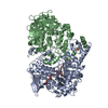
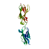
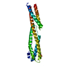
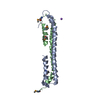
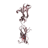
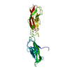
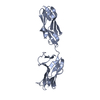
Method to determine structure: MOLECULAR REPLACEMENT
Starting model: POLYALA OF RSBSC AA31-844 STRUCTURE

Resolution: 2.8→24.93 Å / Cor.coef. Fo:Fc: 0.914 / Cor.coef. Fo:Fc free: 0.875 / SU B: 15.101 / SU ML: 0.298 / Cross valid method: THROUGHOUT / ESU R: 0.782 / ESU R Free: 0.378 / Stereochemistry target values: MAXIMUM LIKELIHOOD
Details: HYDROGENS HAVE BEEN ADDED IN THE RIDING POSITIONS. U VALUES REFINED INDIVIDUALLY. HYDROGENS HAVE BEEN USED IF PRESENT IN THE INPUT. CHAIN B CONTAINS DISORDERED REGIONS IN DOMAIN 4
RfactorNum. reflection% reflectionSelection details
Rfree0.28636 684 5 %RANDOM
Rwork0.223 ---
obs0.2262 13044 99.63 %-
Solvent computationIon probe radii: 0.8 Å / Shrinkage radii: 0.8 Å / VDW probe radii: 1.2 Å / Solvent model: MASK
Displacement parametersBiso mean: 40.26 Å2
Baniso -1Baniso -2Baniso -3
1--0.11 Å20 Å20 Å2
2---0.11 Å20 Å2
3---0.23 Å2
Refinement stepCycle: LAST / Resolution: 2.8→24.93 Å
ProteinNucleic acidLigandSolventTotal
Num. atoms2739 0 0 59 2798
Refine LS restraints
Refine-IDTypeDev idealDev ideal targetNumber
X-RAY DIFFRACTIONr_bond_refined_d0.0110.022767
X-RAY DIFFRACTIONr_bond_other_d
X-RAY DIFFRACTIONr_angle_refined_deg1.5591.9683768
X-RAY DIFFRACTIONr_angle_other_deg
X-RAY DIFFRACTIONr_dihedral_angle_1_deg7.035370
X-RAY DIFFRACTIONr_dihedral_angle_2_deg43.58127.80291
X-RAY DIFFRACTIONr_dihedral_angle_3_deg18.45415472
X-RAY DIFFRACTIONr_dihedral_angle_4_deg
X-RAY DIFFRACTIONr_chiral_restr0.090.2488
X-RAY DIFFRACTIONr_gen_planes_refined0.0050.0211967
X-RAY DIFFRACTIONr_gen_planes_other
X-RAY DIFFRACTIONr_nbd_refined
X-RAY DIFFRACTIONr_nbd_other
X-RAY DIFFRACTIONr_nbtor_refined
X-RAY DIFFRACTIONr_nbtor_other
X-RAY DIFFRACTIONr_xyhbond_nbd_refined
X-RAY DIFFRACTIONr_xyhbond_nbd_other
X-RAY DIFFRACTIONr_metal_ion_refined
X-RAY DIFFRACTIONr_metal_ion_other
X-RAY DIFFRACTIONr_symmetry_vdw_refined
X-RAY DIFFRACTIONr_symmetry_vdw_other
X-RAY DIFFRACTIONr_symmetry_hbond_refined
X-RAY DIFFRACTIONr_symmetry_hbond_other
X-RAY DIFFRACTIONr_symmetry_metal_ion_refined
X-RAY DIFFRACTIONr_symmetry_metal_ion_other
X-RAY DIFFRACTIONr_mcbond_it2.2613.9181495
X-RAY DIFFRACTIONr_mcbond_other
X-RAY DIFFRACTIONr_mcangle_it3.7385.8591860
X-RAY DIFFRACTIONr_mcangle_other
X-RAY DIFFRACTIONr_scbond_it2.8654.2111271
X-RAY DIFFRACTIONr_scbond_other
X-RAY DIFFRACTIONr_scangle_it
X-RAY DIFFRACTIONr_scangle_other
X-RAY DIFFRACTIONr_long_range_B_refined
X-RAY DIFFRACTIONr_long_range_B_other
X-RAY DIFFRACTIONr_rigid_bond_restr
X-RAY DIFFRACTIONr_sphericity_free
X-RAY DIFFRACTIONr_sphericity_bonded
LS refinement shellResolution: 2.8→2.872 Å / Total num. of bins used: 20
RfactorNum. reflection% reflection
Rfree0.303 48 -
Rwork0.254 938 -
obs--100 %

+
About Yorodumi

-
News

-
Feb 9, 2022. New format data for meta-information of EMDB entries

New format data for meta-information of EMDB entries

  • Version 3 of the EMDB header file is now the official format.
  • The previous official version 1.9 will be removed from the archive.

Related info.:EMDB header

External links:wwPDB to switch to version 3 of the EMDB data model

-
Aug 12, 2020. Covid-19 info

Covid-19 info

URL: https://pdbj.org/emnavi/covid19.php

New page: Covid-19 featured information page in EM Navigator.

Related info.:Covid-19 info / Mar 5, 2020. Novel coronavirus structure data

+
Mar 5, 2020. Novel coronavirus structure data

Novel coronavirus structure data

Related info.:Yorodumi Speices / Aug 12, 2020. Covid-19 info

External links:COVID-19 featured content - PDBj / Molecule of the Month (242):Coronavirus Proteases

+
Jan 31, 2019. EMDB accession codes are about to change! (news from PDBe EMDB page)

EMDB accession codes are about to change! (news from PDBe EMDB page)

  • The allocation of 4 digits for EMDB accession codes will soon come to an end. Whilst these codes will remain in use, new EMDB accession codes will include an additional digit and will expand incrementally as the available range of codes is exhausted. The current 4-digit format prefixed with “EMD-” (i.e. EMD-XXXX) will advance to a 5-digit format (i.e. EMD-XXXXX), and so on. It is currently estimated that the 4-digit codes will be depleted around Spring 2019, at which point the 5-digit format will come into force.
  • The EM Navigator/Yorodumi systems omit the EMD- prefix.

Related info.:Q: What is EMD? / ID/Accession-code notation in Yorodumi/EM Navigator

External links:EMDB Accession Codes are Changing Soon! / Contact to PDBj

+
Jul 12, 2017. Major update of PDB

Major update of PDB

  • wwPDB released updated PDB data conforming to the new PDBx/mmCIF dictionary.
  • This is a major update changing the version number from 4 to 5, and with Remediation, in which all the entries are updated.
  • In this update, many items about electron microscopy experimental information are reorganized (e.g. em_software).
  • Now, EM Navigator and Yorodumi are based on the updated data.

External links:wwPDB Remediation / Enriched Model Files Conforming to OneDep Data Standards Now Available in the PDB FTP Archive

-
Yorodumi

Thousand views of thousand structures

  • Yorodumi is a browser for structure data from EMDB, PDB, SASBDB, etc.
  • This page is also the successor to EM Navigator detail page, and also detail information page/front-end page for Omokage search.
  • The word "yorodu" (or yorozu) is an old Japanese word meaning "ten thousand". "mi" (miru) is to see.

Related info.:EMDB / PDB / SASBDB / Comparison of 3 databanks / Yorodumi Search / Aug 31, 2016. New EM Navigator & Yorodumi / Yorodumi Papers / Jmol/JSmol / Function and homology information / Changes in new EM Navigator and Yorodumi

Read more