 Movie
Movie Controller

[English] 日本語
 Yorodumi
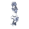
- PDB-2iak: Crystal Structure of a protease resistant fragment of the plakin ... -
+ Open data

- Basic information
| Entry | Database: PDB / ID: 2iak | ||||||
|---|---|---|---|---|---|---|---|
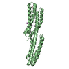
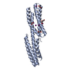
| Title | Crystal Structure of a protease resistant fragment of the plakin domain of Bullous Pemphigoid Antigen1 (BPAG1) | ||||||
 Components Components | Bullous pemphigoid antigen 1, isoform 5 | ||||||
 Keywords Keywords | CELL ADHESION / Triple helical bundle / spectrin repeat | ||||||
| Function / homology |  Function and homology information Function and homology informationtype III intermediate filament / Type I hemidesmosome assembly / neurofilament cytoskeleton / RHOV GTPase cycle / RND3 GTPase cycle / RHOU GTPase cycle / hemidesmosome assembly / hemidesmosome / RND2 GTPase cycle / RND1 GTPase cycle ...type III intermediate filament / Type I hemidesmosome assembly / neurofilament cytoskeleton / RHOV GTPase cycle / RND3 GTPase cycle / RHOU GTPase cycle / hemidesmosome assembly / hemidesmosome / RND2 GTPase cycle / RND1 GTPase cycle / H zone / perinuclear endoplasmic reticulum / intermediate filament cytoskeleton organization / microtubule plus-end / maintenance of cell polarity / microtubule plus-end binding / intermediate filament cytoskeleton / retrograde axonal transport / intermediate filament / cell leading edge / intercalated disc / intracellular transport / regulation of microtubule polymerization or depolymerization / stress fiber / cytoplasmic microtubule organization / cytoskeleton organization / axon cytoplasm / axonogenesis / wound healing / cell motility / sarcolemma / response to wounding / integrin binding / microtubule cytoskeleton organization / Z disc / cytoplasmic side of plasma membrane / nuclear envelope / actin cytoskeleton / microtubule cytoskeleton / actin binding / cytoplasmic vesicle / cell cortex / microtubule binding / cell adhesion / postsynaptic density / axon / focal adhesion / calcium ion binding / endoplasmic reticulum membrane / perinuclear region of cytoplasm / structural molecule activity / protein homodimerization activity / nucleoplasm / membrane / nucleus / cytoplasm / cytosol Similarity search - Function | ||||||
| Biological species |  | ||||||
| Method |  X-RAY DIFFRACTION / SYNCHROTRON / SAD / Resolution: 3 Å X-RAY DIFFRACTION / SYNCHROTRON / SAD / Resolution: 3 Å | ||||||
 Authors Authors | Jefferson, J.J. | ||||||
 Citation Citation | Journal: J.Mol.Biol. / Year: 2007 Title: Structural analysis of the plakin domain of bullous pemphigoid antigen1 (BPAG1) suggests that plakins are members of the spectrin superfamily. Authors: Jefferson, J.J. / Ciatto, C. / Shapiro, L. / Liem, R.K. | ||||||
| History |
|
- Structure visualization
| Structure viewer | Molecule: MolmilJmol/JSmol |
|---|
- Downloads & links
-Download
| PDBx/mmCIF format | 2iak.cif.gz | 49 KB | Display | PDBx/mmCIF format |
|---|---|---|---|---|
| PDB format | pdb2iak.ent.gz | 35.4 KB | Display | PDB format |
| PDBx/mmJSON format | 2iak.json.gz | Tree view | PDBx/mmJSON format | |
| Others |  Other downloads Other downloads |
-Validation report
| Arichive directory | https://data.pdbj.org/pub/pdb/validation_reports/ia/2iakftp://data.pdbj.org/pub/pdb/validation_reports/ia/2iak | HTTPS FTP |
|---|
-Related structure data
| Similar structure data |
|---|


-Links
 PDBj
PDBj- Assembly
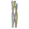
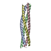
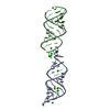
| Deposited unit | 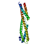
| ||||||||
|---|---|---|---|---|---|---|---|---|---|
| 1 |
| ||||||||
| 2 | x 6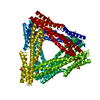
| ||||||||
| 3 | 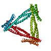
| ||||||||
| Unit cell |
|
-Components
| #1: Protein | Mass: 26559.941 Da / Num. of mol.: 1 / Fragment: Plakin Domain, residues 226-449 / Mutation: V163A Source method: isolated from a genetically manipulated source Source: (gene. exp.)  | ||
|---|---|---|---|
| #2: Chemical |   Mass: 96.063 Da / Num. of mol.: 2 / Source method: obtained synthetically / Formula: SO4 Mass: 96.063 Da / Num. of mol.: 2 / Source method: obtained synthetically / Formula: SO4#3: Water | ChemComp-HOH / |  Mass: 18.015 Da / Num. of mol.: 11 / Source method: isolated from a natural source / Formula: H2O Mass: 18.015 Da / Num. of mol.: 11 / Source method: isolated from a natural source / Formula: H2O |
-Experimental details
-Experiment
| Experiment | Method: X-RAY DIFFRACTION / Number of used crystals: 1 |
|---|
- Sample preparation
| Crystal | Density Matthews: 5.89 Å3/Da / Density % sol: 79.11 % |
|---|---|
| Crystal grow | Temperature: 273 K / Method: vapor diffusion, hanging drop / pH: 9.2 Details: 1.6M ammonium sulphate, 50mM CAPSO, pH 9.2, VAPOR DIFFUSION, HANGING DROP, temperature 273K |
-Data collection
| Diffraction source | Source: SYNCHROTRON / Site: NSLS  / Beamline: X29A / Wavelength: 0.9791 Å / Beamline: X29A / Wavelength: 0.9791 Å |
|---|---|
| Detector | Type: ADSC QUANTUM 315 / Detector: CCD / Date: Oct 27, 2005 |
| Radiation | Protocol: SINGLE WAVELENGTH / Monochromatic (M) / Laue (L): M / Scattering type: x-ray |
| Radiation wavelength | Wavelength: 0.9791 Å / Relative weight: 1 |
| Reflection | Resolution: 3→20 Å / Num. all: 24211 / Num. obs: 24299 / % possible obs: 99.6 % / Observed criterion σ(I): -3 |
| Reflection shell | Resolution: 3→3.18 Å / % possible all: 99.6 |
- Processing
| Software |
| ||||||||||||||||||||||||||||||||||||||||||||||||||||||||||||||||||||||||||||||||||||||||||||||||||||||||||||||||||||||||||||||||||||||||||||||||||||||||||||||||||||||||||
|---|---|---|---|---|---|---|---|---|---|---|---|---|---|---|---|---|---|---|---|---|---|---|---|---|---|---|---|---|---|---|---|---|---|---|---|---|---|---|---|---|---|---|---|---|---|---|---|---|---|---|---|---|---|---|---|---|---|---|---|---|---|---|---|---|---|---|---|---|---|---|---|---|---|---|---|---|---|---|---|---|---|---|---|---|---|---|---|---|---|---|---|---|---|---|---|---|---|---|---|---|---|---|---|---|---|---|---|---|---|---|---|---|---|---|---|---|---|---|---|---|---|---|---|---|---|---|---|---|---|---|---|---|---|---|---|---|---|---|---|---|---|---|---|---|---|---|---|---|---|---|---|---|---|---|---|---|---|---|---|---|---|---|---|---|---|---|---|---|---|---|---|
| Refinement | Method to determine structure: SAD / Resolution: 3→20 Å / Cor.coef. Fo:Fc: 0.918 / Cor.coef. Fo:Fc free: 0.885 / SU B: 24.914 / SU ML: 0.211 / TLS residual ADP flag: LIKELY RESIDUAL / Cross valid method: THROUGHOUT / ESU R: 0.313 / ESU R Free: 0.29 / Stereochemistry target values: MAXIMUM LIKELIHOOD / Details: HYDROGENS HAVE BEEN ADDED IN THE RIDING POSITIONS
| ||||||||||||||||||||||||||||||||||||||||||||||||||||||||||||||||||||||||||||||||||||||||||||||||||||||||||||||||||||||||||||||||||||||||||||||||||||||||||||||||||||||||||
| Solvent computation | Ion probe radii: 0.8 Å / Shrinkage radii: 0.8 Å / VDW probe radii: 1.2 Å / Solvent model: MASK | ||||||||||||||||||||||||||||||||||||||||||||||||||||||||||||||||||||||||||||||||||||||||||||||||||||||||||||||||||||||||||||||||||||||||||||||||||||||||||||||||||||||||||
| Displacement parameters | Biso mean: 41.221 Å2 | ||||||||||||||||||||||||||||||||||||||||||||||||||||||||||||||||||||||||||||||||||||||||||||||||||||||||||||||||||||||||||||||||||||||||||||||||||||||||||||||||||||||||||
| Refinement step | Cycle: LAST / Resolution: 3→20 Å
| ||||||||||||||||||||||||||||||||||||||||||||||||||||||||||||||||||||||||||||||||||||||||||||||||||||||||||||||||||||||||||||||||||||||||||||||||||||||||||||||||||||||||||
| Refine LS restraints |
| ||||||||||||||||||||||||||||||||||||||||||||||||||||||||||||||||||||||||||||||||||||||||||||||||||||||||||||||||||||||||||||||||||||||||||||||||||||||||||||||||||||||||||
| LS refinement shell | Highest resolution: 3 Å / Num. reflection Rwork: 892 / Total num. of bins used: 20 | ||||||||||||||||||||||||||||||||||||||||||||||||||||||||||||||||||||||||||||||||||||||||||||||||||||||||||||||||||||||||||||||||||||||||||||||||||||||||||||||||||||||||||
| Refinement TLS params. | Method: refined / Refine-ID: X-RAY DIFFRACTION
| ||||||||||||||||||||||||||||||||||||||||||||||||||||||||||||||||||||||||||||||||||||||||||||||||||||||||||||||||||||||||||||||||||||||||||||||||||||||||||||||||||||||||||
| Refinement TLS group |
|
