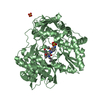
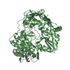
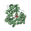
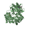
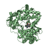
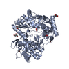
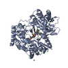
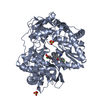
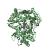
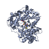
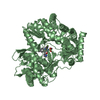
Entry Database : PDB / ID : 4tn2Title NS5b in complex with lactam-thiophene carboxylic acids Genome polyprotein Keywords / Function / homology Function Domain/homology Component
/ / / / / / / / / / / / / / / / / / / / / / / / / / / / / / / / / / / / / / / / / / / / / / / / / / / / / / / / / / / / / / / / / / / / / / / / / / / / / / / / / / / / / / / / / / / / / / / / / / / / / / / / / / / / / Biological species Method / / Resolution : 2.7 Å Authors Chopra, R. Journal : Bioorg.Med.Chem.Lett. / Year : 2014Title : Design and synthesis of lactam-thiophene carboxylic acids as potent hepatitis C virus polymerase inhibitors.Authors : Barnes-Seeman, D. / Boiselle, C. / Capacci-Daniel, C. / Chopra, R. / Hoffmaster, K. / Jones, C.T. / Kato, M. / Lin, K. / Ma, S. / Pan, G. / Shu, L. / Wang, J. / Whiteman, L. / Xu, M. / Zheng, R. / Fu, J. History Deposition Jun 2, 2014 Deposition site / Processing site Revision 1.0 Sep 17, 2014 Provider / Type Revision 1.1 Feb 4, 2015 Group Revision 1.2 Jun 1, 2016 Group Revision 1.3 Nov 22, 2017 Group / Refinement description / Category / softwareItem / _software.classification / _software.nameRevision 1.4 Dec 27, 2023 Group / Database referencesCategory chem_comp_atom / chem_comp_bond ... chem_comp_atom / chem_comp_bond / database_2 / diffrn_radiation_wavelength Item / _database_2.pdbx_database_accession
Show all Show less
 Movie
Movie Controller


 Open data
 Basic information
 Components
 Keywords
 Function and homology information
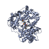
 Hepatitis B virus
 X-RAY DIFFRACTION /
  Authors
 Citation
 Structure visualization
 Downloads & links
 Other downloads


 PDBj
PDBj
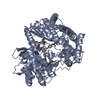
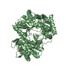
 Assembly
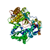
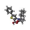
 Mass: 369.477 Da / Num. of mol.: 1 / Source method: obtained synthetically / Formula: C21H23NO3S
 Mass: 18.015 Da / Num. of mol.: 127 / Source method: isolated from a natural source / Formula: H2O
 Sample preparation
 Processing