- PDB-4rgl: Crystal structure of a Fic family protein (Dde_2494) from Desulfo... -
+
Open data
ID or keywords:
Loading...
-
Basic information
Entry
Database: PDB / ID: 4rgl
Title
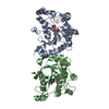
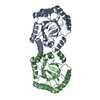
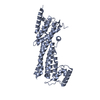
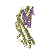
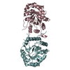
Crystal structure of a Fic family protein (Dde_2494) from Desulfovibrio desulfuricans G20 at 2.70 A resolution
Components
Filamentation induced by cAMP protein Fic
Keywords
DNA BINDING PROTEIN / PF02661 family / Fic/DOC protein / Structural Genomics / Joint Center for Structural Genomics / JCSG / Protein Structure Initiative / PSI-BIOLOGY
Mass: 18.015 Da / Num. of mol.: 20 / Source method: isolated from a natural source / Formula: H2O
Has protein modification
Y
Sequence details
THE CONSTRUCT (1-342) WAS EXPRESSED WITH A PURIFICATION TAG MGSDKIHHHHHHENLYFQG. THE TAG WAS ...THE CONSTRUCT (1-342) WAS EXPRESSED WITH A PURIFICATION TAG MGSDKIHHHHHHENLYFQG. THE TAG WAS REMOVED WITH TEV PROTEASE LEAVING ONLY A GLYCINE (0) FOLLOWED BY THE TARGET SEQUENCE.
-
Experimental details
-
Experiment
Experiment
Method: X-RAY DIFFRACTION / Number of used crystals: 1
-
Sample preparation
Crystal
Density Matthews: 2.52 Å3/Da / Density % sol: 51.18 %
Crystal grow
Temperature: 277 K / Method: vapor diffusion, sitting drop / pH: 10.5 Details: 40.0% MPD, 0.1M CAPS pH 10.5, NANODROP, VAPOR DIFFUSION, SITTING DROP, temperature 277K
Type: MARMOSAIC 325 mm CCD / Detector: CCD / Date: Apr 7, 2007 Details: Flat mirror (vertical focusing); single crystal Si(111) bent monochromator (horizontal focusing)
Radiation
Monochromator: single crystal Si(111) bent / Protocol: MAD / Monochromatic (M) / Laue (L): M / Scattering type: x-ray
Radiation wavelength
ID
Wavelength (Å)
Relative weight
1
0.97895
1
2
0.97929
1
3
0.91837
1
Reflection
Resolution: 2.7→41.908 Å / Num. obs: 11879 / % possible obs: 99.3 % / Observed criterion σ(I): -3 / Redundancy: 6.83 % / Biso Wilson estimate: 79.008 Å2 / Rmerge(I) obs: 0.086 / Net I/σ(I): 13.33
Reflection shell
Diffraction-ID: 1
Resolution (Å)
Redundancy (%)
Rmerge(I) obs
Mean I/σ(I) obs
Num. measured obs
Num. unique obs
% possible all
2.7-2.8
6.88
1.261
1.7
7724
1123
94.8
2.8-2.91
0.831
2.6
7987
1125
100
2.91-3.04
0.584
3.6
8007
1129
100
3.04-3.2
0.364
5.3
8292
1168
100
3.2-3.4
0.22
7.8
8227
1171
100
3.4-3.66
0.144
11.1
8035
1168
100
3.66-4.03
0.086
16.6
8198
1202
99.8
4.03-4.6
0.058
23.1
8142
1179
100
4.6-5.77
0.053
26
8229
1230
99.9
5.77-41.91
0.045
30.4
8287
1384
98.8
-
Phasing
Phasing
Method: MAD
-
Processing
Software
Name
Version
Classification
NB
MolProbity
3beta29
modelbuilding
PDB_EXTRACT
3.1
dataextraction
SHELX
phasing
SHARP
phasing
XSCALE
January10, 2014BUILT=20140307
datascaling
BUSTER-TNT
2.10.0
refinement
XDS
datareduction
SHELXD
phasing
BUSTER
2.10.0
refinement
Refinement
Method to determine structure: MAD / Resolution: 2.7→41.908 Å / Cor.coef. Fo:Fc: 0.9201 / Cor.coef. Fo:Fc free: 0.8767 / Occupancy max: 1 / Occupancy min: 0.75 / Cross valid method: THROUGHOUT / σ(F): 0 Details: 1. A MET-INHIBITION PROTOCOL WAS USED FOR SELENOMETHIONINE INCORPORATION DURING PROTEIN EXPRESSION. THE OCCUPANCY OF THE SE ATOMS IN THE MSE RESIDUES WAS REDUCED TO 0.75 FOR THE REDUCED ...Details: 1. A MET-INHIBITION PROTOCOL WAS USED FOR SELENOMETHIONINE INCORPORATION DURING PROTEIN EXPRESSION. THE OCCUPANCY OF THE SE ATOMS IN THE MSE RESIDUES WAS REDUCED TO 0.75 FOR THE REDUCED SCATTERING POWER DUE TO PARTIAL S-MET INCORPORATION. 2. ATOM RECORD CONTAINS SUM OF TLS AND RESIDUAL B FACTORS. ANISOU RECORD CONTAINS SUM OF TLS AND RESIDUAL U FACTORS. 3. THE MAD PHASES WERE USED AS RESTRAINTS DURING REFINEMENT.4. VAL 33 IS IN A REGIONS OF POOR ELECTRON DENSITY AND IS RAMACHANDRAN OUTLIER IN MOLPROBITY. 5.ELECTRON DENSITY CORRESPONDING TO AN INTER-DOMAIN LINKER BETWEEN THR 257-GLN 263 IS DISORDERED AND THIS REGION COULD NOT BE RELIABLY MODELED.THE POSITIONING OF THE TWO DOMAINS FROM A SINGLE SUBUNIT IS BASED ON THE PROXIMITY OF THE C-TERMINAL END OF ONE DOMAIN TO THE N-TERMINAL END OF THE SECOND DOMAIN. 6. AN UNKNOWN LIGAND (UNL) HAS BEEN MODELED INTO THE PUTATIVE ACTIVE SITE.
In the structure databanks used in Yorodumi, some data are registered as the other names, "COVID-19 virus" and "2019-nCoV". Here are the details of the virus and the list of structure data.
Jan 31, 2019. EMDB accession codes are about to change! (news from PDBe EMDB page)
EMDB accession codes are about to change! (news from PDBe EMDB page)
The allocation of 4 digits for EMDB accession codes will soon come to an end. Whilst these codes will remain in use, new EMDB accession codes will include an additional digit and will expand incrementally as the available range of codes is exhausted. The current 4-digit format prefixed with “EMD-” (i.e. EMD-XXXX) will advance to a 5-digit format (i.e. EMD-XXXXX), and so on. It is currently estimated that the 4-digit codes will be depleted around Spring 2019, at which point the 5-digit format will come into force.
The EM Navigator/Yorodumi systems omit the EMD- prefix.
Related info.:Q: What is EMD? / ID/Accession-code notation in Yorodumi/EM Navigator
Yorodumi is a browser for structure data from EMDB, PDB, SASBDB, etc.
This page is also the successor to EM Navigator detail page, and also detail information page/front-end page for Omokage search.
The word "yorodu" (or yorozu) is an old Japanese word meaning "ten thousand". "mi" (miru) is to see.
Related info.:EMDB / PDB / SASBDB / Comparison of 3 databanks / Yorodumi Search / Aug 31, 2016. New EM Navigator & Yorodumi / Yorodumi Papers / Jmol/JSmol / Function and homology information / Changes in new EM Navigator and Yorodumi
 Movie
Movie Controller

 Yorodumi
 Open data
 Basic information
 Components
 Keywords
 Function and homology information
 Desulfovibrio alaskensis (bacteria)
 X-RAY DIFFRACTION /
  Authors
 Citation
 Structure visualization
 Downloads & links
 Other downloads
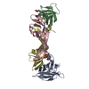


 PDBj
PDBj

 Assembly
 Mass: 18.015 Da / Num. of mol.: 20 / Source method: isolated from a natural source / Formula: H2O
 Sample preparation
 / Beamline: BL11-1 / Wavelength: 0.97895,0.97929,0.91837
 Processing