 Movie
Movie Controller


+ Open data

- Basic information
| Entry | Database: PDB / ID: 4rex | ||||||
|---|---|---|---|---|---|---|---|
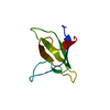
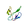
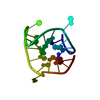
| Title | Crystal structure of the first WW domain of human YAP2 isoform | ||||||
 Components Components | Yorkie homolog | ||||||
 Keywords Keywords |  PROTEIN BINDING / WW domain / Hippo signaling pathway / antiparallel beta-sheet / proline rich motifs PROTEIN BINDING / WW domain / Hippo signaling pathway / antiparallel beta-sheet / proline rich motifs | ||||||
| Function / homology |  Function and homology information Function and homology informationenterocyte differentiation / regulation of keratinocyte proliferation / regulation of metanephric nephron tubule epithelial cell differentiation / cardiac muscle tissue regeneration / contact inhibition / glandular epithelial cell differentiation / TEAD-YAP complex / lateral mesoderm development / RUNX3 regulates YAP1-mediated transcription / bud elongation involved in lung branching ...enterocyte differentiation / regulation of keratinocyte proliferation / regulation of metanephric nephron tubule epithelial cell differentiation / cardiac muscle tissue regeneration / contact inhibition / glandular epithelial cell differentiation / TEAD-YAP complex / lateral mesoderm development / RUNX3 regulates YAP1-mediated transcription / bud elongation involved in lung branching / polarized epithelial cell differentiation / notochord development / negative regulation of cilium assembly / lung epithelial cell differentiation / YAP1- and WWTR1 (TAZ)-stimulated gene expression / heart process / trophectodermal cell differentiation / paraxial mesoderm development / tissue homeostasis / hippo signaling / regulation of stem cell proliferation / EGR2 and SOX10-mediated initiation of Schwann cell myelination / intestinal epithelial cell development / negative regulation of epithelial cell apoptotic process / Formation of axial mesoderm / negative regulation of stem cell differentiation / embryonic heart tube morphogenesis / female germ cell nucleus / proline-rich region binding / Signaling by Hippo / positive regulation of intracellular estrogen receptor signaling pathway / negative regulation of epithelial cell differentiation / negative regulation of fat cell differentiation / organ growth / positive regulation of stem cell population maintenance / interleukin-6-mediated signaling pathway / positive regulation of Notch signaling pathway / RUNX2 regulates osteoblast differentiation / progesterone receptor signaling pathway / Zygotic genome activation (ZGA) / somatic stem cell population maintenance / regulation of neurogenesis / canonical Wnt signaling pathway / positive regulation of osteoblast differentiation / vasculogenesis / Nuclear signaling by ERBB4 / cellular response to retinoic acid / extrinsic apoptotic signaling pathway / keratinocyte differentiation / positive regulation of cardiac muscle cell proliferation / response to progesterone / epithelial cell proliferation / positive regulation of epithelial cell proliferation / negative regulation of extrinsic apoptotic signaling pathway / cellular response to gamma radiation / wound healing / cell morphogenesis / positive regulation of protein localization to nucleus / positive regulation of canonical Wnt signaling pathway / transcription corepressor activity / cell-cell junction / cell junction / RUNX1 regulates transcription of genes involved in differentiation of HSCs / positive regulation of cell growth / protein-containing complex assembly / DNA-binding transcription factor binding / cell population proliferation / transcription coactivator activity / transcription cis-regulatory region binding / RNA polymerase II cis-regulatory region sequence-specific DNA binding / negative regulation of gene expression / DNA damage response / chromatin binding / positive regulation of gene expression / positive regulation of DNA-templated transcription / negative regulation of transcription by RNA polymerase II / positive regulation of transcription by RNA polymerase II / nucleoplasm / membrane / nucleus / cytosol / cytoplasmSimilarity search - Function | ||||||
| Biological species |  Homo sapiens (human) Homo sapiens (human) | ||||||
| Method | X-RAY DIFFRACTION / SYNCHROTRON / MOLECULAR REPLACEMENT / molecular replacement / Resolution: 1.6 Å | ||||||
 Authors Authors | Camara-Artigas, A. | ||||||
 Citation Citation | Journal: J.Struct.Biol. / Year: 2015 Title: Crystal structure of the first WW domain of human YAP2 isoform. Authors: Martinez-Rodriguez, S. / Bacarizo, J. / Luque, I. / Camara-Artigas, A. | ||||||
| History |
|
- Structure visualization
| Structure viewer | Molecule: MolmilJmol/JSmol |
|---|
- Downloads & links
-Download
| PDBx/mmCIF format | 4rex.cif.gz | 41.1 KB | Display | PDBx/mmCIF format |
|---|---|---|---|---|
| PDB format | pdb4rex.ent.gz | 28.5 KB | Display | PDB format |
| PDBx/mmJSON format | 4rex.json.gz | Tree view | PDBx/mmJSON format | |
| Others |  Other downloads Other downloads |
-Validation report
| Arichive directory | https://data.pdbj.org/pub/pdb/validation_reports/re/4rexftp://data.pdbj.org/pub/pdb/validation_reports/re/4rex | HTTPS FTP |
|---|
-Related structure data
| Related structure data |  2ho2S S: Starting model for refinement |
|---|---|
| Similar structure data |


-Links
 PDBj
PDBj


- Assembly
| Deposited unit | 
| ||||||||
|---|---|---|---|---|---|---|---|---|---|
| 1 |
| ||||||||
| Unit cell |
| ||||||||
| Components on special symmetry positions |
|
-Components
| #1: Protein/peptide | Mass: 5529.180 Da / Num. of mol.: 1 / Fragment: WW domain (UNP residues 165-209) Source method: isolated from a genetically manipulated source Source: (gene. exp.) Homo sapiens (human) / Gene: YAP1, YAP65 / Plasmid: pETM30 / Production host:  Escherichia coli (E. coli) / Strain (production host): BL21(DE3) / References: UniProt: P46937 Escherichia coli (E. coli) / Strain (production host): BL21(DE3) / References: UniProt: P46937 |
|---|---|
| #2: Chemical | ChemComp-SO4 / Sulfate  Mass: 96.063 Da / Num. of mol.: 1 / Source method: obtained synthetically / Formula: SO4 Mass: 96.063 Da / Num. of mol.: 1 / Source method: obtained synthetically / Formula: SO4 |
| #3: Water | ChemComp-HOH / Water Mass: 18.015 Da / Num. of mol.: 42 / Source method: isolated from a natural source / Formula: H2O Mass: 18.015 Da / Num. of mol.: 42 / Source method: isolated from a natural source / Formula: H2O |
-Experimental details
-Experiment
| Experiment | Method: X-RAY DIFFRACTION / Number of used crystals: 1 |
|---|
- Sample preparation
| Crystal | Density Matthews: 1.77 Å3/Da / Density % sol: 30.57 % |
|---|---|
| Crystal grow | Temperature: 293 K / pH: 5 Details: 1.5 M ammonium sulphate, 0.1 M sodium acetate, 100 mM proline, 5 mM NDSB-201 and 5mM MBCD, pH 5.0, VAPOR DIFFUSION, HANGING DROP, temperature 293K |
-Data collection
| Diffraction | Mean temperature: 100 K | |||||||||||||||
|---|---|---|---|---|---|---|---|---|---|---|---|---|---|---|---|---|
| Diffraction source | Source: SYNCHROTRON / Site: ALBA  / Beamline: XALOC / Wavelength: 0.97926 / Beamline: XALOC / Wavelength: 0.97926 | |||||||||||||||
| Detector | Type: DECTRIS PILATUS 6M / Detector: PIXEL / Date: Oct 24, 2013 Details: SI(111) CHANNEL-CUT CRYSTAL MONOCHROMATOR AND A PAIR OF KB MIRRORS | |||||||||||||||
| Radiation | Monochromator: SI(111) CHANNEL-CUT CRYSTAL MONOCHROMATOR / Protocol: SINGLE WAVELENGTH / Monochromatic (M) / Laue (L): M / Scattering type: x-ray | |||||||||||||||
| Radiation wavelength | Wavelength: 0.97926 Å / Relative weight: 1 | |||||||||||||||
| Reflection | Redundancy: 6.9 % / Number: 38300 / Rmerge(I) obs: 0.104 / D res high: 1.6 Å / D res low: 42.67 Å / Num. obs: 5514 / % possible obs: 99.2 / Rejects: 10 | |||||||||||||||
| Diffraction reflection shell |
| |||||||||||||||
| Reflection | Resolution: 1.6→42.67 Å / Num. obs: 5514 / % possible obs: 99.2 % / Observed criterion σ(I): 0 / Redundancy: 6.9 % / Biso Wilson estimate: 12.78 Å2 / Rmerge(I) obs: 0.104 / Rsym value: 0.104 / Net I/σ(I): 12 | |||||||||||||||
| Reflection shell | Resolution: 1.6→1.63 Å / Redundancy: 6.8 % / Rmerge(I) obs: 0.582 / Mean I/σ(I) obs: 3.2 / % possible all: 96.9 |
-Phasing
| Phasing | Method: molecular replacement |
|---|
- Processing
| Software |
| |||||||||||||||||||||||||||||||||||||||||||||||||||||||||||||||||||||||||||||||||||||||||||||||||||||||||||||||||||||||||||||
|---|---|---|---|---|---|---|---|---|---|---|---|---|---|---|---|---|---|---|---|---|---|---|---|---|---|---|---|---|---|---|---|---|---|---|---|---|---|---|---|---|---|---|---|---|---|---|---|---|---|---|---|---|---|---|---|---|---|---|---|---|---|---|---|---|---|---|---|---|---|---|---|---|---|---|---|---|---|---|---|---|---|---|---|---|---|---|---|---|---|---|---|---|---|---|---|---|---|---|---|---|---|---|---|---|---|---|---|---|---|---|---|---|---|---|---|---|---|---|---|---|---|---|---|---|---|---|
| Refinement | Method to determine structure: MOLECULAR REPLACEMENT Starting model: 2HO2 Resolution: 1.6→21.551 Å / SU ML: 0.16 / σ(F): 0.47 / Phase error: 23.42 / Stereochemistry target values: ML
| |||||||||||||||||||||||||||||||||||||||||||||||||||||||||||||||||||||||||||||||||||||||||||||||||||||||||||||||||||||||||||||
| Solvent computation | Shrinkage radii: 0.9 Å / VDW probe radii: 1.11 Å / Solvent model: FLAT BULK SOLVENT MODEL | |||||||||||||||||||||||||||||||||||||||||||||||||||||||||||||||||||||||||||||||||||||||||||||||||||||||||||||||||||||||||||||
| Displacement parameters | Biso mean: 16.7 Å2 | |||||||||||||||||||||||||||||||||||||||||||||||||||||||||||||||||||||||||||||||||||||||||||||||||||||||||||||||||||||||||||||
| Refinement step | Cycle: LAST / Resolution: 1.6→21.551 Å
| |||||||||||||||||||||||||||||||||||||||||||||||||||||||||||||||||||||||||||||||||||||||||||||||||||||||||||||||||||||||||||||
| Refine LS restraints |
| |||||||||||||||||||||||||||||||||||||||||||||||||||||||||||||||||||||||||||||||||||||||||||||||||||||||||||||||||||||||||||||
| LS refinement shell |
| |||||||||||||||||||||||||||||||||||||||||||||||||||||||||||||||||||||||||||||||||||||||||||||||||||||||||||||||||||||||||||||
| Refinement TLS params. | Method: refined / Refine-ID: X-RAY DIFFRACTION
| |||||||||||||||||||||||||||||||||||||||||||||||||||||||||||||||||||||||||||||||||||||||||||||||||||||||||||||||||||||||||||||
| Refinement TLS group |
|
