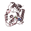
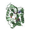
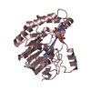
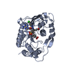
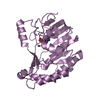
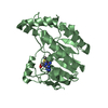
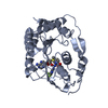
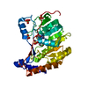
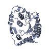
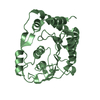
[English] 日本語
Yorodumi
- PDB-4r05: Crystal structure of the refolded DENV3 methyltransferase -

+
Open data


ID or keywords:

Loading...

-
Basic information

Entry
Database: PDB / ID: 4r05
TitleCrystal structure of the refolded DENV3 methyltransferase
ComponentsNonstructural protein NS5
KeywordsTRANSFERASE / METHYLTRANSFERASE / flavivirus
Function / homology
Function and homology information


symbiont-mediated suppression of host JAK-STAT cascade via inhibition of host TYK2 activity / host cell mitochondrion / symbiont-mediated suppression of host JAK-STAT cascade via inhibition of STAT2 activity / symbiont-mediated suppression of host cytoplasmic pattern recognition receptor signaling pathway via inhibition of MAVS activity / ribonucleoside triphosphate phosphatase activity / viral capsid / : / double-stranded RNA binding / protein complex oligomerization / monoatomic ion channel activity ...symbiont-mediated suppression of host JAK-STAT cascade via inhibition of host TYK2 activity / host cell mitochondrion / symbiont-mediated suppression of host JAK-STAT cascade via inhibition of STAT2 activity / symbiont-mediated suppression of host cytoplasmic pattern recognition receptor signaling pathway via inhibition of MAVS activity / ribonucleoside triphosphate phosphatase activity / viral capsid / : / double-stranded RNA binding / protein complex oligomerization / monoatomic ion channel activity / clathrin-dependent endocytosis of virus by host cell / mRNA (nucleoside-2'-O-)-methyltransferase activity / mRNA 5'-cap (guanine-N7-)-methyltransferase activity / RNA helicase activity / host cell endoplasmic reticulum membrane / protein dimerization activity / induction by virus of host autophagy / viral RNA genome replication / RNA-dependent RNA polymerase activity / serine-type endopeptidase activity / fusion of virus membrane with host endosome membrane / viral envelope / symbiont-mediated suppression of host type I interferon-mediated signaling pathway / host cell nucleus / structural molecule activity / virion attachment to host cell / virion membrane / proteolysis / extracellular region / ATP binding / membrane / metal ion binding
Similarity search - Function
: / : / Flavivirus envelope glycoprotein E, stem/anchor domain / RNA-directed RNA polymerase, thumb domain, Flavivirus / Flavivirus RNA-directed RNA polymerase, thumb domain / Flavivirus capsid protein C superfamily / Flavivirus non-structural protein NS2B / Flavivirus NS3 helicase, C-terminal helical domain / Genome polyprotein, Flavivirus / Flavivirus non-structural protein NS4A ...: / : / Flavivirus envelope glycoprotein E, stem/anchor domain / RNA-directed RNA polymerase, thumb domain, Flavivirus / Flavivirus RNA-directed RNA polymerase, thumb domain / Flavivirus capsid protein C superfamily / Flavivirus non-structural protein NS2B / Flavivirus NS3 helicase, C-terminal helical domain / Genome polyprotein, Flavivirus / Flavivirus non-structural protein NS4A / Flavivirus non-structural protein NS2B / Flavivirus capsid protein C / Flavivirus non-structural protein NS4B / mRNA cap 0/1 methyltransferase / Flavivirus capsid protein C / Flavivirus non-structural protein NS4B / Flavivirus non-structural protein NS4A / Flavivirus NS2B domain profile. / mRNA cap 0 and cap 1 methyltransferase (EC 2.1.1.56 and EC 2.1.1.57) domain profile. / Flavivirus non-structural protein NS2A / Flavivirus non-structural protein NS2A / Flavivirus NS3, petidase S7 / Peptidase S7, Flavivirus NS3 serine protease / Flavivirus NS3 protease (NS3pro) domain profile. / Envelope glycoprotein M, flavivirus / Flavivirus envelope glycoprotein M / RNA-directed RNA polymerase, flavivirus / Flavivirus RNA-directed RNA polymerase, fingers and palm domains / Flavivirus non-structural Protein NS1 / Flavivirus non-structural protein NS1 / Envelope glycoprotein M superfamily, flavivirus / Flavivirus polyprotein propeptide / Flavivirus polyprotein propeptide superfamily / Flavivirus polyprotein propeptide / Flaviviral glycoprotein E, central domain, subdomain 1 / Flaviviral glycoprotein E, central domain, subdomain 2 / Flavivirus envelope glycoprotein E, Stem/Anchor domain / Flavivirus glycoprotein E, immunoglobulin-like domain / Flavivirus envelope glycoprotein E, Stem/Anchor domain superfamily / Flavivirus glycoprotein, immunoglobulin-like domain / Flavivirus glycoprotein central and dimerisation domain / Flavivirus glycoprotein, central and dimerisation domains / Ribosomal RNA methyltransferase, FtsJ domain / FtsJ-like methyltransferase / Flavivirus/Alphavirus glycoprotein, immunoglobulin-like domain superfamily / Flavivirus glycoprotein, central and dimerisation domain superfamily / Flaviviral glycoprotein E, dimerisation domain / DEAD box, Flavivirus / Flavivirus DEAD domain / Vaccinia Virus protein VP39 / helicase superfamily c-terminal domain / Immunoglobulin E-set / Superfamilies 1 and 2 helicase C-terminal domain profile. / Superfamilies 1 and 2 helicase ATP-binding type-1 domain profile. / DEAD-like helicases superfamily / Helicase, C-terminal / Helicase superfamily 1/2, ATP-binding domain / RNA-directed RNA polymerase, catalytic domain / RdRp of positive ssRNA viruses catalytic domain profile. / S-adenosyl-L-methionine-dependent methyltransferase superfamily / Peptidase S1, PA clan, chymotrypsin-like fold / Peptidase S1, PA clan / DNA/RNA polymerase superfamily / P-loop containing nucleoside triphosphate hydrolase / Rossmann fold / 3-Layer(aba) Sandwich / Alpha Beta
Similarity search - Domain/homology
Biological speciesDengue virus 3
MethodX-RAY DIFFRACTION / MOLECULAR REPLACEMENT / Resolution: 2.1 Å
AuthorsBrecher, M.B. / Li, Z. / Zhang, J. / Chen, H. / Lin, Q. / Liu, B. / Li, H.M.
CitationJournal: Protein Sci. / Year: 2015
Title: Refolding of a fully functional flavivirus methyltransferase revealed that S-adenosyl methionine but not S-adenosyl homocysteine is copurified with flavivirus methyltransferase.
Authors: Brecher, M.B. / Li, Z. / Zhang, J. / Chen, H. / Lin, Q. / Liu, B. / Li, H.
History
DepositionJul 29, 2014Deposition site: RCSB / Processing site: RCSB
Revision 1.0Nov 12, 2014Provider: repository / Type: Initial release
Revision 1.1Jan 21, 2015Group: Database references
Revision 1.2Sep 20, 2023Group: Data collection / Database references / Refinement description
Category: chem_comp_atom / chem_comp_bond ...chem_comp_atom / chem_comp_bond / database_2 / pdbx_initial_refinement_model / struct_ref_seq_dif
Item: _database_2.pdbx_DOI / _database_2.pdbx_database_accession / _struct_ref_seq_dif.details

-
Structure visualization

Structure viewerMolecule:
MolmilJmol/JSmol

Downloads & links

-
Assembly

Deposited unit
A: Nonstructural protein NS5
B: Nonstructural protein NS5


Theoretical massNumber of molelcules
Total (without water)60,1392
Polymers60,1392
Non-polymers00
Water4,468248
1
A: Nonstructural protein NS5


Theoretical massNumber of molelcules
Total (without water)30,0701
Polymers30,0701
Non-polymers00
Water181
TypeNameSymmetry operationNumber
identity operation1_555x,y,z1
MethodPISA
2
B: Nonstructural protein NS5


Theoretical massNumber of molelcules
Total (without water)30,0701
Polymers30,0701
Non-polymers00
Water181
TypeNameSymmetry operationNumber
identity operation1_555x,y,z1
MethodPISA
Unit cell
Length a, b, c (Å)44.688, 48.386, 68.073
Angle α, β, γ (deg.)79.11, 78.75, 69.71
Int Tables number1
Space group name H-MP1

-
Components

#1: Protein Nonstructural protein NS5


Mass: 30069.586 Da / Num. of mol.: 2 / Fragment: UNP residues 2491-2752
Source method: isolated from a genetically manipulated source
Source: (gene. exp.) Dengue virus 3 / Gene: NS5 / Plasmid: pET28a-3C / Production host: Escherichia coli (E. coli) / Strain (production host): BL21(DE3) / References: UniProt: C1KBQ3
#2: Water ChemComp-HOH / water / Water


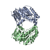
Mass: 18.015 Da / Num. of mol.: 248 / Source method: isolated from a natural source / Formula: H2O

-
Experimental details

-
Experiment

ExperimentMethod: X-RAY DIFFRACTION / Number of used crystals: 1

-
Sample preparation

CrystalDensity Matthews: 2.23 Å3/Da / Density % sol: 44.89 %
Crystal growTemperature: 298 K / Method: vapor diffusion, hanging drop / pH: 5.6
Details: 50 mM sodium citrate, pH 5.6, 24-30% PEG 4,000, 5% saturated ammonium sulfate, 10% glycerol, 0-20% DMSO, and 5 mM DTT, VAPOR DIFFUSION, HANGING DROP, temperature 298K

-
Data collection

DiffractionMean temperature: 100 K
Diffraction sourceSource: ROTATING ANODE / Type: RIGAKU MICROMAX-007 / Wavelength: 1.5418 Å
DetectorType: RIGAKU RAXIS IV++ / Detector: IMAGE PLATE / Date: May 13, 2013
RadiationMonochromator: YALE MIRRORS / Protocol: SINGLE WAVELENGTH / Monochromatic (M) / Laue (L): M / Scattering type: x-ray
Radiation wavelengthWavelength: 1.5418 Å / Relative weight: 1
ReflectionResolution: 2.1→45 Å / Num. all: 28779 / Num. obs: 28779 / % possible obs: 94.9 % / Observed criterion σ(F): 1 / Observed criterion σ(I): 1 / Redundancy: 2.1 % / Rmerge(I) obs: 0.123 / Rsym value: 0.123 / Net I/σ(I): 3.9
Reflection shellResolution: 2.1→2.18 Å / Redundancy: 2.1 % / Rmerge(I) obs: 0.45 / Mean I/σ(I) obs: 1.3 / Num. unique all: 2579 / Rsym value: 0.45 / % possible all: 92.7

-
Processing

Software
NameVersionClassification
CrystalCleardata collection
PHASERphasing
PHENIX(phenix.refine: 1.8.4_1496)refinement
CrystalCleardata reduction
CrystalCleardata scaling
RefinementMethod to determine structure: MOLECULAR REPLACEMENT
Starting model: 3P8Z
Resolution: 2.1→33.092 Å / SU ML: 0.4 / Cross valid method: THROUGHOUT / σ(F): 1.96 / σ(I): 1 / Phase error: 37.49 / Stereochemistry target values: ML
RfactorNum. reflection% reflectionSelection details
Rfree0.3072 1479 5.15 %RANDOM
Rwork0.2496 ---
obs0.2526 28706 94.65 %-
all-28779 --
Solvent computationShrinkage radii: 0.9 Å / VDW probe radii: 1.11 Å / Solvent model: FLAT BULK SOLVENT MODEL
Refine analyzeLuzzati coordinate error obs: 0.4 Å
Refinement stepCycle: LAST / Resolution: 2.1→33.092 Å
ProteinNucleic acidLigandSolventTotal
Num. atoms4094 0 0 248 4342
Refine LS restraints
Refine-IDTypeDev idealNumber
X-RAY DIFFRACTIONf_bond_d0.0044182
X-RAY DIFFRACTIONf_angle_d0.8215638
X-RAY DIFFRACTIONf_dihedral_angle_d13.4321610
X-RAY DIFFRACTIONf_chiral_restr0.033608
X-RAY DIFFRACTIONf_plane_restr0.004720
LS refinement shell
Resolution (Å)Rfactor RfreeNum. reflection RfreeRfactor RworkNum. reflection RworkRefine-ID% reflection obs (%)
2.1-2.16780.39931270.40452393X-RAY DIFFRACTION92
2.1678-2.24530.42281320.38662462X-RAY DIFFRACTION93
2.2453-2.33510.40821310.33992423X-RAY DIFFRACTION93
2.3351-2.44140.39581330.32542485X-RAY DIFFRACTION94
2.4414-2.570.35311390.31132442X-RAY DIFFRACTION94
2.57-2.7310.33931240.28422495X-RAY DIFFRACTION95
2.731-2.94170.31921410.26722520X-RAY DIFFRACTION96
2.9417-3.23750.30141360.24732505X-RAY DIFFRACTION96
3.2375-3.70550.28511370.20932521X-RAY DIFFRACTION97
3.7055-4.66640.23131460.18662493X-RAY DIFFRACTION96
4.6664-33.09570.30391330.21842488X-RAY DIFFRACTION95

+
About Yorodumi

-
News

-
Feb 9, 2022. New format data for meta-information of EMDB entries

New format data for meta-information of EMDB entries

  • Version 3 of the EMDB header file is now the official format.
  • The previous official version 1.9 will be removed from the archive.

Related info.:EMDB header

External links:wwPDB to switch to version 3 of the EMDB data model

-
Aug 12, 2020. Covid-19 info

Covid-19 info

URL: https://pdbj.org/emnavi/covid19.php

New page: Covid-19 featured information page in EM Navigator.

Related info.:Covid-19 info / Mar 5, 2020. Novel coronavirus structure data

+
Mar 5, 2020. Novel coronavirus structure data

Novel coronavirus structure data

Related info.:Yorodumi Speices / Aug 12, 2020. Covid-19 info

External links:COVID-19 featured content - PDBj / Molecule of the Month (242):Coronavirus Proteases

+
Jan 31, 2019. EMDB accession codes are about to change! (news from PDBe EMDB page)

EMDB accession codes are about to change! (news from PDBe EMDB page)

  • The allocation of 4 digits for EMDB accession codes will soon come to an end. Whilst these codes will remain in use, new EMDB accession codes will include an additional digit and will expand incrementally as the available range of codes is exhausted. The current 4-digit format prefixed with “EMD-” (i.e. EMD-XXXX) will advance to a 5-digit format (i.e. EMD-XXXXX), and so on. It is currently estimated that the 4-digit codes will be depleted around Spring 2019, at which point the 5-digit format will come into force.
  • The EM Navigator/Yorodumi systems omit the EMD- prefix.

Related info.:Q: What is EMD? / ID/Accession-code notation in Yorodumi/EM Navigator

External links:EMDB Accession Codes are Changing Soon! / Contact to PDBj

+
Jul 12, 2017. Major update of PDB

Major update of PDB

  • wwPDB released updated PDB data conforming to the new PDBx/mmCIF dictionary.
  • This is a major update changing the version number from 4 to 5, and with Remediation, in which all the entries are updated.
  • In this update, many items about electron microscopy experimental information are reorganized (e.g. em_software).
  • Now, EM Navigator and Yorodumi are based on the updated data.

External links:wwPDB Remediation / Enriched Model Files Conforming to OneDep Data Standards Now Available in the PDB FTP Archive

-
Yorodumi

Thousand views of thousand structures

  • Yorodumi is a browser for structure data from EMDB, PDB, SASBDB, etc.
  • This page is also the successor to EM Navigator detail page, and also detail information page/front-end page for Omokage search.
  • The word "yorodu" (or yorozu) is an old Japanese word meaning "ten thousand". "mi" (miru) is to see.

Related info.:EMDB / PDB / SASBDB / Comparison of 3 databanks / Yorodumi Search / Aug 31, 2016. New EM Navigator & Yorodumi / Yorodumi Papers / Jmol/JSmol / Function and homology information / Changes in new EM Navigator and Yorodumi

Read more