 Movie
Movie Controller

[English] 日本語
 Yorodumi
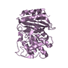
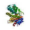
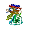
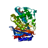
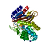
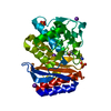
- PDB-4qy5: Crystal structures of chimeric beta-lactamase cTEM-19m showing di... -
+ Open data

- Basic information
| Entry | Database: PDB / ID: 4qy5 | ||||||
|---|---|---|---|---|---|---|---|
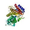
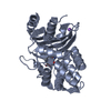
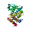
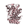
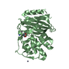
| Title | Crystal structures of chimeric beta-lactamase cTEM-19m showing different conformations | ||||||
 Components Components | Beta-lactamase TEM,Beta-lactamase PSE-4 | ||||||
 Keywords Keywords | HYDROLASE | ||||||
| Function / homology |  Function and homology information Function and homology informationbeta-lactam antibiotic catabolic process / beta-lactamase activity / beta-lactamase / response to antibiotic Similarity search - Function | ||||||
| Biological species |   Pseudomonas aeruginosa (bacteria) Pseudomonas aeruginosa (bacteria) | ||||||
| Method |  X-RAY DIFFRACTION / SYNCHROTRON / MOLECULAR REPLACEMENT / Resolution: 1.501 Å X-RAY DIFFRACTION / SYNCHROTRON / MOLECULAR REPLACEMENT / Resolution: 1.501 Å | ||||||
 Authors Authors | Park, J. / Gobeil, S. / Pelletier, J.N. / Berghuis, A.M. | ||||||
 Citation Citation | Journal: To be Published Title: Crystal structures of chimeric beta-lactamase cTEM-19m showing different conformations Authors: Park, J. / Gobeil, S. / Pelletier, J.N. / Berghuis, A.M. | ||||||
| History |
|
- Structure visualization
| Structure viewer | Molecule: MolmilJmol/JSmol |
|---|
- Downloads & links
-Download
| PDBx/mmCIF format | 4qy5.cif.gz | 136 KB | Display | PDBx/mmCIF format |
|---|---|---|---|---|
| PDB format | pdb4qy5.ent.gz | 104.6 KB | Display | PDB format |
| PDBx/mmJSON format | 4qy5.json.gz | Tree view | PDBx/mmJSON format | |
| Others |  Other downloads Other downloads |
-Validation report
| Summary document | 4qy5_validation.pdf.gz | 409.5 KB | Display | wwPDB validaton report |
|---|---|---|---|---|
| Full document | 4qy5_full_validation.pdf.gz | 409.7 KB | Display | |
| Data in XML | 4qy5_validation.xml.gz | 14.6 KB | Display | |
| Data in CIF | 4qy5_validation.cif.gz | 23 KB | Display | |
| Arichive directory | https://data.pdbj.org/pub/pdb/validation_reports/qy/4qy5ftp://data.pdbj.org/pub/pdb/validation_reports/qy/4qy5 | HTTPS FTP |
-Related structure data
| Related structure data | 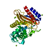 4qy6C  4zg4S S: Starting model for refinement C: citing same article ( |
|---|---|
| Similar structure data |


-Links
 PDBj
PDBj


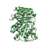
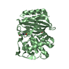
- Assembly
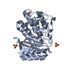
| Deposited unit | 
| ||||||||
|---|---|---|---|---|---|---|---|---|---|
| 1 |
| ||||||||
| Unit cell |
|
-Components
| #1: Protein | Mass: 28827.830 Da / Num. of mol.: 1 Source method: isolated from a genetically manipulated source Source: (gene. exp.) Pseudomonas aeruginosa (bacteria)Gene: bla, blaT-3, blaT-4, blaT-5, blaT-6, pse4, carB1 / Production host: References: UniProt: P62593, UniProt: P16897, beta-lactamase | ||||
|---|---|---|---|---|---|
| #2: Chemical | ChemComp-CL /   Mass: 35.453 Da / Num. of mol.: 4 / Source method: obtained synthetically / Formula: Cl Mass: 35.453 Da / Num. of mol.: 4 / Source method: obtained synthetically / Formula: Cl#3: Chemical |   Mass: 24.305 Da / Num. of mol.: 3 / Source method: obtained synthetically / Formula: Mg Mass: 24.305 Da / Num. of mol.: 3 / Source method: obtained synthetically / Formula: Mg#4: Water | ChemComp-HOH / |  Mass: 18.015 Da / Num. of mol.: 317 / Source method: isolated from a natural source / Formula: H2O Mass: 18.015 Da / Num. of mol.: 317 / Source method: isolated from a natural source / Formula: H2O |
-Experimental details
-Experiment
| Experiment | Method: X-RAY DIFFRACTION / Number of used crystals: 1 |
|---|
- Sample preparation
| Crystal | Density Matthews: 2.02 Å3/Da / Density % sol: 39.07 % |
|---|---|
| Crystal grow | Temperature: 295 K / Method: vapor diffusion, sitting drop / pH: 8.5 Details: 30% PEG 4000, 0.2M magnesium chloride, 0.1M TrisHCl, pH 8.5, VAPOR DIFFUSION, SITTING DROP, temperature 295K |
-Data collection
| Diffraction | Mean temperature: 100 K |
|---|---|
| Diffraction source | Source: SYNCHROTRON / Site: CLSI  / Beamline: 08ID-1 / Wavelength: 0.97949 Å / Beamline: 08ID-1 / Wavelength: 0.97949 Å |
| Detector | Type: RAYONIX MX-300 / Detector: CCD / Date: Aug 12, 2011 |
| Radiation | Monochromator: ACCEL/BRUKER DCM / Protocol: SINGLE WAVELENGTH / Monochromatic (M) / Laue (L): M / Scattering type: x-ray |
| Radiation wavelength | Wavelength: 0.97949 Å / Relative weight: 1 |
| Reflection | Resolution: 1.5→51.4 Å / Num. all: 38195 / Num. obs: 38195 / % possible obs: 99.9 % / Redundancy: 7.2 % / Biso Wilson estimate: 12.3 Å2 / Rmerge(I) obs: 0.062 / Net I/σ(I): 20.9 |
| Reflection shell | Resolution: 1.5→1.54 Å / Redundancy: 7.1 % / Rmerge(I) obs: 0.841 / Mean I/σ(I) obs: 2.2 / Num. unique all: 2767 / % possible all: 99.3 |
- Processing
| Software |
| ||||||||||||||||||||||||||||||||||||||||||||||||||||||||||||||||||||||||||||||||||||||||||||||||||||||||||||||||||||||||
|---|---|---|---|---|---|---|---|---|---|---|---|---|---|---|---|---|---|---|---|---|---|---|---|---|---|---|---|---|---|---|---|---|---|---|---|---|---|---|---|---|---|---|---|---|---|---|---|---|---|---|---|---|---|---|---|---|---|---|---|---|---|---|---|---|---|---|---|---|---|---|---|---|---|---|---|---|---|---|---|---|---|---|---|---|---|---|---|---|---|---|---|---|---|---|---|---|---|---|---|---|---|---|---|---|---|---|---|---|---|---|---|---|---|---|---|---|---|---|---|---|---|
| Refinement | Method to determine structure: MOLECULAR REPLACEMENT Starting model: PDB ENTRY 4ZG4 Resolution: 1.501→51.4 Å / Cor.coef. Fo:Fc: 0.984 / Cor.coef. Fo:Fc free: 0.971 / SU B: 2.834 / SU ML: 0.046 / Cross valid method: THROUGHOUT / ESU R: 0.071 / ESU R Free: 0.064 / Stereochemistry target values: MAXIMUM LIKELIHOOD / Details: HYDROGENS HAVE BEEN ADDED IN THE RIDING POSITIONS
| ||||||||||||||||||||||||||||||||||||||||||||||||||||||||||||||||||||||||||||||||||||||||||||||||||||||||||||||||||||||||
| Solvent computation | Ion probe radii: 0.8 Å / Shrinkage radii: 0.8 Å / VDW probe radii: 1.2 Å / Solvent model: MASK | ||||||||||||||||||||||||||||||||||||||||||||||||||||||||||||||||||||||||||||||||||||||||||||||||||||||||||||||||||||||||
| Displacement parameters | Biso mean: 18.022 Å2
| ||||||||||||||||||||||||||||||||||||||||||||||||||||||||||||||||||||||||||||||||||||||||||||||||||||||||||||||||||||||||
| Refinement step | Cycle: LAST / Resolution: 1.501→51.4 Å
| ||||||||||||||||||||||||||||||||||||||||||||||||||||||||||||||||||||||||||||||||||||||||||||||||||||||||||||||||||||||||
| Refine LS restraints |
| ||||||||||||||||||||||||||||||||||||||||||||||||||||||||||||||||||||||||||||||||||||||||||||||||||||||||||||||||||||||||
| LS refinement shell | Resolution: 1.501→1.54 Å / Total num. of bins used: 20
|
