 Movie
Movie Controller

[English] 日本語
 Yorodumi
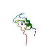
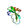
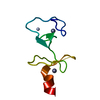
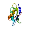
- PDB-4pti: THE GEOMETRY OF THE REACTIVE SITE AND OF THE PEPTIDE GROUPS IN TR... -
+ Open data

- Basic information
| Entry | Database: PDB / ID: 4pti | |||||||||
|---|---|---|---|---|---|---|---|---|---|---|
| Title | THE GEOMETRY OF THE REACTIVE SITE AND OF THE PEPTIDE GROUPS IN TRYPSIN, TRYPSINOGEN AND ITS COMPLEXES WITH INHIBITORS | |||||||||
 Components Components | TRYPSIN INHIBITOR | |||||||||
 Keywords Keywords | PROTEINASE INHIBITOR (TRYPSIN) | |||||||||
| Function / homology |  Function and homology information Function and homology informationtrypsinogen activation / negative regulation of serine-type endopeptidase activity / sulfate binding / potassium channel inhibitor activity / negative regulation of platelet aggregation / zymogen binding / molecular function inhibitor activity / negative regulation of thrombin-activated receptor signaling pathway / serine protease inhibitor complex / serine-type endopeptidase inhibitor activity ...trypsinogen activation / negative regulation of serine-type endopeptidase activity / sulfate binding / potassium channel inhibitor activity / negative regulation of platelet aggregation / zymogen binding / molecular function inhibitor activity / negative regulation of thrombin-activated receptor signaling pathway / serine protease inhibitor complex / serine-type endopeptidase inhibitor activity / protease binding / calcium ion binding / extracellular spaceSimilarity search - Function | |||||||||
| Biological species |  Bos taurus (cattle) Bos taurus (cattle) | |||||||||
| Method | X-RAY DIFFRACTION / Resolution: 1.5 Å | |||||||||
 Authors Authors | Huber, R. / Kukla, D. / Ruehlmann, A. / Epp, O. / Formanek, H. / Deisenhofer, J. / Steigemann, W. | |||||||||
 Citation Citation | Journal: Acta Crystallogr.,Sect.B / Year: 1983 Title: The Geometry of the Reactive Site and of the Peptide Groups in Trypsin, Trypsinogen and its Complexes with Inhibitors Authors: Marquart, M. / Walter, J. / Deisenhofer, J. / Bode, W. / Huber, R. #1: Journal: J.Mol.Biol. / Year: 1987Title: Comparison of Two Highly Refined Structures of Bovine Pancreatic Trypsin Inhibitor Authors: Wlodawer, A. / Deisenhofer, J. / Huber, R. #2: Journal: Acta Crystallogr.,Sect.B / Year: 1975Title: Crystallographic Refinement of the Structure of Bovine Pancreatic Trypsin Inhibitor at 1.5 Angstroms Resolution Authors: Deisenhofer, J. / Steigemann, W. #3: Journal: Bayer Symp. / Year: 1974Title: The Model of the Basic Pancreatic Trypsin Inhibitor Refined at 1.5 Angstroms Resolution Authors: Deisenhofer, J. / Steigemann, W. #4: Journal: Cold Spring Harbor Symp.Quant.Biol. / Year: 1972Title: Pancreatic Trypsin Inhibitor (Kunitz). Part I. Structure and Function Authors: Huber, R. / Kukla, D. / Ruehlmann, A. / Steigemann, W. #5: Journal: Cold Spring Harbor Symp.Quant.Biol. / Year: 1972Title: Pancreatic Trypsin Inhibitor (Kunitz). Part II. Complexes with Proteinases Authors: Ruehlmann, A. / Schramm, H.J. / Kukla, D. / Huber, R. #6: Journal: Naturwissenschaften / Year: 1970Title: The Basic Trypsin Inhibitor of Bovine Pancreas. I. Structure Analysis and Conformation of the Polypeptide Chain Authors: Huber, R. / Kukla, D. / Ruehlmann, A. / Epp, O. / Formanek, H. | |||||||||
| History |
|
- Structure visualization
| Structure viewer | Molecule: MolmilJmol/JSmol |
|---|
- Downloads & links
-Download
| PDBx/mmCIF format | 4pti.cif.gz | 19.3 KB | Display | PDBx/mmCIF format |
|---|---|---|---|---|
| PDB format | pdb4pti.ent.gz | 15.2 KB | Display | PDB format |
| PDBx/mmJSON format | 4pti.json.gz | Tree view | PDBx/mmJSON format | |
| Others |  Other downloads Other downloads |
-Validation report
| Arichive directory | https://data.pdbj.org/pub/pdb/validation_reports/pt/4ptiftp://data.pdbj.org/pub/pdb/validation_reports/pt/4pti | HTTPS FTP |
|---|
-Related structure data
| Similar structure data |
|---|


-Links
 PDBj
PDBj


- Assembly
| Deposited unit | 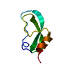
| ||||||||
|---|---|---|---|---|---|---|---|---|---|
| 1 |
| ||||||||
| Unit cell |
|
-Components
| #1: Protein | Mass: 6527.568 Da / Num. of mol.: 1 Source method: isolated from a genetically manipulated source Source: (gene. exp.) Bos taurus (cattle) / References: UniProt: P00974 |
|---|---|
| #2: Water | ChemComp-HOH / Water Mass: 18.015 Da / Num. of mol.: 60 / Source method: isolated from a natural source / Formula: H2O Mass: 18.015 Da / Num. of mol.: 60 / Source method: isolated from a natural source / Formula: H2O |
-Experimental details
-Experiment
| Experiment | Method: X-RAY DIFFRACTION |
|---|
- Sample preparation
| Crystal | Density Matthews: 1.84 Å3/Da / Density % sol: 33.01 % |
|---|---|
| Crystal grow | *PLUS Method: unknown |
- Processing
| Refinement | Resolution: 1.5→7 Å / Rfactor Rwork: 0.162 | ||||||||||||
|---|---|---|---|---|---|---|---|---|---|---|---|---|---|
| Refinement step | Cycle: LAST / Resolution: 1.5→7 Å
|
