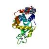
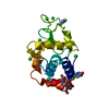
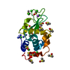
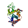
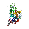
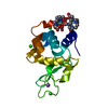
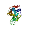
Entry Database : PDB / ID : 4n5rTitle Hen egg-white lysozyme phased using free-electron laser data Lysozyme C Keywords / Function / homology Function Domain/homology Component
/ / / / / / / / / / / / / / / / / / / / / / / / / / / / / / / / / / / / / / / / / / / Biological species Gallus gallus (chicken)Method / / / Resolution : 2.1 Å Authors Barends, T.R.M. / Foucar, L. / Botha, S. / Doak, R.B. / Shoeman, R.L. / Nass, K. / Koglin, J.E. / Williams, G.J. / Boutet, S. / Messerschmidt, M. / Schlichting, I. Journal : Nature / Year : 2014Title : De novo protein crystal structure determination from X-ray free-electron laser data.Authors : Barends, T.R. / Foucar, L. / Botha, S. / Doak, R.B. / Shoeman, R.L. / Nass, K. / Koglin, J.E. / Williams, G.J. / Boutet, S. / Messerschmidt, M. / Schlichting, I. History Deposition Oct 10, 2013 Deposition site / Processing site Revision 1.0 Nov 27, 2013 Provider / Type Revision 1.1 Dec 25, 2013 Group Revision 1.2 Jan 1, 2014 Group Revision 1.3 Jan 22, 2014 Group Revision 1.4 Feb 14, 2018 Group / Category Item / _diffrn_source.pdbx_synchrotron_siteRevision 1.5 Aug 16, 2023 Group / Database references / Derived calculationsCategory chem_comp_atom / chem_comp_bond ... chem_comp_atom / chem_comp_bond / database_2 / pdbx_related_exp_data_set / struct_conn / struct_site Item _database_2.pdbx_DOI / _database_2.pdbx_database_accession ... _database_2.pdbx_DOI / _database_2.pdbx_database_accession / _struct_conn.pdbx_dist_value / _struct_conn.ptnr1_auth_seq_id / _struct_conn.ptnr1_label_asym_id / _struct_conn.ptnr2_auth_seq_id / _struct_conn.ptnr2_label_asym_id / _struct_conn.ptnr2_label_atom_id / _struct_site.pdbx_auth_asym_id / _struct_site.pdbx_auth_comp_id / _struct_site.pdbx_auth_seq_id Revision 1.6 Sep 20, 2023 Group / Category Revision 1.7 Nov 6, 2024 Group / Category / pdbx_modification_feature
Show all Show less
 Movie
Movie Controller


 Open data
 Basic information
 Components
 Keywords
 Function and homology information
 X-RAY DIFFRACTION /
  Authors
 Citation
 Structure visualization
 Downloads & links
 Other downloads
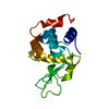

 PDBj
PDBj


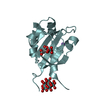
 Assembly
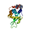
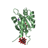
 Mass: 157.250 Da / Num. of mol.: 2 / Source method: obtained synthetically / Formula: Gd
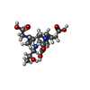
 Mass: 404.459 Da / Num. of mol.: 2 / Source method: obtained synthetically / Formula: C17H32N4O7
 Mass: 18.015 Da / Num. of mol.: 52 / Source method: isolated from a natural source / Formula: H2O
 Sample preparation
 / Beamline: CXI / Wavelength: 1.45 Å
 Processing