 Movie
Movie Controller

[English] 日本語
 Yorodumi
- PDB-4mm0: Crystal Structure Analysis of the Putative Thioether Synthase Sgv... -
+ Open data

- Basic information
| Entry | Database: PDB / ID: 4mm0 | ||||||
|---|---|---|---|---|---|---|---|
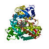
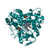
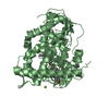
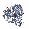
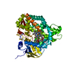
| Title | Crystal Structure Analysis of the Putative Thioether Synthase SgvP Involved in the Tailoring Step of Griseoviridin | ||||||
 Components Components | P450-like monooxygenase | ||||||
 Keywords Keywords |  OXIDOREDUCTASE / Cytochrome P450 / monooxygenase OXIDOREDUCTASE / Cytochrome P450 / monooxygenase | ||||||
| Function / homology |  Function and homology information Function and homology informationoxidoreductase activity, acting on paired donors, with incorporation or reduction of molecular oxygen / monooxygenase activity / iron ion binding / heme bindingSimilarity search - Function | ||||||
| Biological species |  Streptomyces griseoviridis (bacteria) Streptomyces griseoviridis (bacteria) | ||||||
| Method | X-RAY DIFFRACTION / SYNCHROTRON / MOLECULAR REPLACEMENT / Resolution: 2.6 Å | ||||||
 Authors Authors | Zhang, H. / Huang, L. / Yi, M. / Cai, T. / Zhang, H. | ||||||
 Citation Citation | Journal: To be Published Title: Structural Analysis of SgvP: a Putative Thioether Synthase Involved in the Tailoring Step of Griseoviridin Authors: Zhang, H. / Huang, L. / Yi, M. / Cai, T. / Zhang, H. | ||||||
| History |
|
- Structure visualization
| Structure viewer | Molecule: MolmilJmol/JSmol |
|---|
- Downloads & links
-Download
| PDBx/mmCIF format | 4mm0.cif.gz | 172 KB | Display | PDBx/mmCIF format |
|---|---|---|---|---|
| PDB format | pdb4mm0.ent.gz | 134.9 KB | Display | PDB format |
| PDBx/mmJSON format | 4mm0.json.gz | Tree view | PDBx/mmJSON format | |
| Others |  Other downloads Other downloads |
-Validation report
| Arichive directory | https://data.pdbj.org/pub/pdb/validation_reports/mm/4mm0ftp://data.pdbj.org/pub/pdb/validation_reports/mm/4mm0 | HTTPS FTP |
|---|
-Related structure data
| Related structure data |  2y46S S: Starting model for refinement |
|---|---|
| Similar structure data |

-Links
 PDBj
PDBj
- Assembly
| Deposited unit | 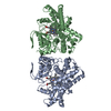
| ||||||||
|---|---|---|---|---|---|---|---|---|---|
| 1 | 
| ||||||||
| 2 | 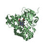
| ||||||||
| Unit cell |
|
-Components
| #1: Protein | Mass: 43903.902 Da / Num. of mol.: 2 Source method: isolated from a genetically manipulated source Source: (gene. exp.) Streptomyces griseoviridis (bacteria) / Plasmid: pET28a / Production host: Escherichia coli (E. coli) / Strain (production host): BL21(DE3)References: UniProt: R9USI6*PLUS, Oxidoreductases; Acting on paired donors, with incorporation or reduction of molecular oxygen; With NADH or NADPH as one donor, and incorporation of one atom of ...References: UniProt: R9USI6*PLUS, Oxidoreductases; Acting on paired donors, with incorporation or reduction of molecular oxygen; With NADH or NADPH as one donor, and incorporation of one atom of oxygen into the other donor#2: Chemical | Heme B  Mass: 616.487 Da / Num. of mol.: 2 / Source method: obtained synthetically / Formula: C34H32FeN4O4 Mass: 616.487 Da / Num. of mol.: 2 / Source method: obtained synthetically / Formula: C34H32FeN4O4#3: Water | ChemComp-HOH / | Water Mass: 18.015 Da / Num. of mol.: 383 / Source method: isolated from a natural source / Formula: H2O Mass: 18.015 Da / Num. of mol.: 383 / Source method: isolated from a natural source / Formula: H2OSequence details | A SEQUENCE DATABASE REFERENCE FOR THIS PROTEIN DOES NOT CURRENTLY EXIST IN UNIPROT. THE SEQUENCE ...A SEQUENCE DATABASE REFERENCE FOR THIS PROTEIN DOES NOT CURRENTLY EXIST IN UNIPROT. THE SEQUENCE HAS BEEN DEPOSITED TO DDBJ WITH ACCESSION NUMBER AGN74891.1. N-TERMINAL RESIDUES MET-ALA-SER ARE FROM EXPRESSION | |
|---|
-Experimental details
-Experiment
| Experiment | Method: X-RAY DIFFRACTION / Number of used crystals: 1 |
|---|
- Sample preparation
| Crystal | Density Matthews: 3.87 Å3/Da / Density % sol: 68.24 % |
|---|---|
| Crystal grow | Temperature: 277.15 K / Method: vapor diffusion, sitting drop / pH: 8.2 Details: 20%(w/v) Polyethylene glycol 3350, 0.2M Potassium sodium tartrate tetrahydrate, 3.0%(v/v) Dimethyl sulfoxide, pH 8.2, VAPOR DIFFUSION, SITTING DROP, temperature 277.15K |
-Data collection
| Diffraction | Mean temperature: 77.15 K |
|---|---|
| Diffraction source | Source: SYNCHROTRON / Site: SSRF  / Beamline: BL17U / Wavelength: 0.979228 Å / Beamline: BL17U / Wavelength: 0.979228 Å |
| Detector | Type: ADSC QUANTUM 315 / Detector: CCD / Date: May 2, 2012 |
| Radiation | Protocol: SINGLE WAVELENGTH / Monochromatic (M) / Laue (L): M / Scattering type: x-ray |
| Radiation wavelength | Wavelength: 0.979228 Å / Relative weight: 1 |
| Reflection | Resolution: 2.6→29.856 Å / Num. all: 43006 / Num. obs: 42958 / % possible obs: 99.89 % / Observed criterion σ(F): 2 / Observed criterion σ(I): 3.11 |
| Reflection shell | Resolution: 2.6→2.67 Å / % possible all: 98.3 |
- Processing
| Software |
| ||||||||||||||||||||||||||||||||||||||||||||||||||||||||||||||||||||||||||||||||||||||||||||||||
|---|---|---|---|---|---|---|---|---|---|---|---|---|---|---|---|---|---|---|---|---|---|---|---|---|---|---|---|---|---|---|---|---|---|---|---|---|---|---|---|---|---|---|---|---|---|---|---|---|---|---|---|---|---|---|---|---|---|---|---|---|---|---|---|---|---|---|---|---|---|---|---|---|---|---|---|---|---|---|---|---|---|---|---|---|---|---|---|---|---|---|---|---|---|---|---|---|---|
| Refinement | Method to determine structure: MOLECULAR REPLACEMENT Starting model: PDB ENTRY 2Y46 Resolution: 2.6→29.856 Å / Occupancy max: 1 / Occupancy min: 1 / SU ML: 0.25 / σ(F): 2.03 / Phase error: 19.62 / Stereochemistry target values: ML
| ||||||||||||||||||||||||||||||||||||||||||||||||||||||||||||||||||||||||||||||||||||||||||||||||
| Solvent computation | Shrinkage radii: 0.9 Å / VDW probe radii: 1.11 Å / Solvent model: FLAT BULK SOLVENT MODEL | ||||||||||||||||||||||||||||||||||||||||||||||||||||||||||||||||||||||||||||||||||||||||||||||||
| Displacement parameters | Biso max: 143.81 Å2 / Biso mean: 30.0195 Å2 / Biso min: 4.95 Å2 | ||||||||||||||||||||||||||||||||||||||||||||||||||||||||||||||||||||||||||||||||||||||||||||||||
| Refinement step | Cycle: LAST / Resolution: 2.6→29.856 Å
| ||||||||||||||||||||||||||||||||||||||||||||||||||||||||||||||||||||||||||||||||||||||||||||||||
| Refine LS restraints |
| ||||||||||||||||||||||||||||||||||||||||||||||||||||||||||||||||||||||||||||||||||||||||||||||||
| LS refinement shell | Refine-ID: X-RAY DIFFRACTION / Total num. of bins used: 15 / % reflection obs: 100 %
|
