satellite DNA binding / pyrimidine-specific mismatch base pair DNA N-glycosylase activity / depyrimidination / DNA N-glycosylase activity / Displacement of DNA glycosylase by APEX1 / Hydrolases; Glycosylases; Hydrolysing N-glycosyl compounds / Recognition and association of DNA glycosylase with site containing an affected pyrimidine / Cleavage of the damaged pyrimidine / DNA endonuclease activity / response to estradiol ...satellite DNA binding / pyrimidine-specific mismatch base pair DNA N-glycosylase activity / depyrimidination / DNA N-glycosylase activity / Displacement of DNA glycosylase by APEX1 / Hydrolases; Glycosylases; Hydrolysing N-glycosyl compounds / Recognition and association of DNA glycosylase with site containing an affected pyrimidine / Cleavage of the damaged pyrimidine / DNA endonuclease activity / response to estradiol / nuclear speck / DNA repair / DNA binding / nucleoplasm / nucleus Similarity search - Function
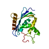
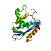
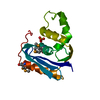
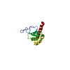
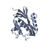
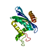
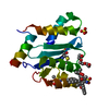
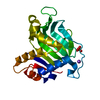
Methyl-CpG-binding domain protein 4 / Methyl-cpg-binding Protein 2; Chain A / Methyl-cpg-binding Protein 2; Chain A / Methyl-CpG binding protein MeCP2/MBD4 / DNA glycosylase / Methyl-CpG binding domain / Methyl-CpG DNA binding / Methyl-CpG binding domain / Methyl-CpG-binding domain (MBD) profile. / DNA-binding domain superfamily ...Methyl-CpG-binding domain protein 4 / Methyl-cpg-binding Protein 2; Chain A / Methyl-cpg-binding Protein 2; Chain A / Methyl-CpG binding protein MeCP2/MBD4 / DNA glycosylase / Methyl-CpG binding domain / Methyl-CpG DNA binding / Methyl-CpG binding domain / Methyl-CpG-binding domain (MBD) profile. / DNA-binding domain superfamily / 2-Layer Sandwich / Alpha Beta Similarity search - Domain/homology
A: Methyl-CpG-binding domain protein 4 B: DNA (5'-D(*GP*CP*CP*AP*AP*(5CM)P*GP*TP*TP*GP*GP*C)-3') C: DNA (5'-D(*GP*CP*CP*AP*AP*(5CM)P*GP*TP*TP*GP*GP*C)-3')
Resolution: 2.5→36 Å / Cor.coef. Fo:Fc: 0.9332 / Cor.coef. Fo:Fc free: 0.9179 / Occupancy max: 1 / Occupancy min: 1 / SU R Cruickshank DPI: 0.38 / Cross valid method: THROUGHOUT / σ(F): 0 Details: Refmac, phenix, coot, molprobity server were also used during refinement. We note the small number of reflections in the x-validation free set and a significant discrepancy between average ...Details: Refmac, phenix, coot, molprobity server were also used during refinement. We note the small number of reflections in the x-validation free set and a significant discrepancy between average refined and Wilson B-factors.
In the structure databanks used in Yorodumi, some data are registered as the other names, "COVID-19 virus" and "2019-nCoV". Here are the details of the virus and the list of structure data.
Jan 31, 2019. EMDB accession codes are about to change! (news from PDBe EMDB page)
EMDB accession codes are about to change! (news from PDBe EMDB page)
The allocation of 4 digits for EMDB accession codes will soon come to an end. Whilst these codes will remain in use, new EMDB accession codes will include an additional digit and will expand incrementally as the available range of codes is exhausted. The current 4-digit format prefixed with “EMD-” (i.e. EMD-XXXX) will advance to a 5-digit format (i.e. EMD-XXXXX), and so on. It is currently estimated that the 4-digit codes will be depleted around Spring 2019, at which point the 5-digit format will come into force.
The EM Navigator/Yorodumi systems omit the EMD- prefix.
Related info.:Q: What is EMD? / ID/Accession-code notation in Yorodumi/EM Navigator
Yorodumi is a browser for structure data from EMDB, PDB, SASBDB, etc.
This page is also the successor to EM Navigator detail page, and also detail information page/front-end page for Omokage search.
The word "yorodu" (or yorozu) is an old Japanese word meaning "ten thousand". "mi" (miru) is to see.
Related info.:EMDB / PDB / SASBDB / Comparison of 3 databanks / Yorodumi Search / Aug 31, 2016. New EM Navigator & Yorodumi / Yorodumi Papers / Jmol/JSmol / Function and homology information / Changes in new EM Navigator and Yorodumi
 Movie
Movie Controller

 Yorodumi
 Open data
 Basic information
 Components
 Keywords
 Function and homology information
 Homo sapiens (human)
 X-RAY DIFFRACTION /
  Authors
 Citation
 Structure visualization
 Downloads & links
 Other downloads
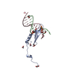
 PDBj
PDBj


 Assembly


 Num. of mol.: 11 / Source method: obtained synthetically
 Sample preparation
 Processing