 Movie
Movie Controller


+ Open data

- Basic information
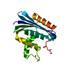
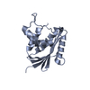
| Entry | Database: PDB / ID: 1kvb | ||||||
|---|---|---|---|---|---|---|---|
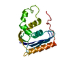
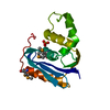
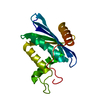
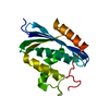
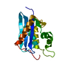
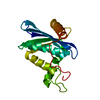
| Title | E. COLI RIBONUCLEASE HI D134H MUTANT | ||||||
 Components Components | RIBONUCLEASE H | ||||||
 Keywords Keywords | ENDORIBONUCLEASE / HYDROLASE / MUTANT | ||||||
| Function / homology |  Function and homology information Function and homology informationDNA replication, removal of RNA primer / ribonuclease H / RNA-DNA hybrid ribonuclease activity / endonuclease activity / nucleic acid binding / magnesium ion binding / cytoplasm Similarity search - Function | ||||||
| Biological species |  | ||||||
| Method |  X-RAY DIFFRACTION / Resolution: 1.9 Å X-RAY DIFFRACTION / Resolution: 1.9 Å | ||||||
 Authors Authors | Kashiwagi, T. / Jeanteur, D. / Haruki, M. / Katayanagi, K. / Kanaya, S. / Morikawa, K. | ||||||
 Citation Citation | Journal: Protein Eng. / Year: 1996 Title: Proposal for new catalytic roles for two invariant residues in Escherichia coli ribonuclease HI. Authors: Kashiwagi, T. / Jeanteur, D. / Haruki, M. / Katayanagi, K. / Kanaya, S. / Morikawa, K. #1: Journal: Proteins / Year: 1993Title: Crystal Structure of Escherichia Coli Rnase Hi in Complex with Mg2+ at 2.8 A Resolution: Proof for a Single Mg(2+)-Binding Site Authors: Katayanagi, K. / Okumura, M. / Morikawa, K. #2: Journal: J.Mol.Biol. / Year: 1992Title: Structural Details of Ribonuclease H from Escherichia Coli as Refined to an Atomic Resolution Authors: Katayanagi, K. / Miyagawa, M. / Matsushima, M. / Ishikawa, M. / Kanaya, S. / Nakamura, H. / Ikehara, M. / Matsuzaki, T. / Morikawa, K. #3: Journal: Science / Year: 1990Title: Structure of Ribonuclease H Phased at 2 A Resolution by MAD Analysis of the Selenomethionyl Protein Authors: Yang, W. / Hendrickson, W.A. / Crouch, R.J. / Satow, Y. #4: Journal: Nature / Year: 1990Title: Three-Dimensional Structure of Ribonuclease H from E. Coli Authors: Katayanagi, K. / Miyagawa, M. / Matsushima, M. / Ishikawa, M. / Kanaya, S. / Ikehara, M. / Matsuzaki, T. / Morikawa, K. | ||||||
| History |
|
- Structure visualization
| Structure viewer | Molecule: MolmilJmol/JSmol |
|---|
- Downloads & links
-Download
| PDBx/mmCIF format | 1kvb.cif.gz | 47.1 KB | Display | PDBx/mmCIF format |
|---|---|---|---|---|
| PDB format | pdb1kvb.ent.gz | 33.5 KB | Display | PDB format |
| PDBx/mmJSON format | 1kvb.json.gz | Tree view | PDBx/mmJSON format | |
| Others |  Other downloads Other downloads |
-Validation report
| Arichive directory | https://data.pdbj.org/pub/pdb/validation_reports/kv/1kvbftp://data.pdbj.org/pub/pdb/validation_reports/kv/1kvb | HTTPS FTP |
|---|
-Related structure data


-Links
 PDBj
PDBj
- Assembly
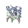
| Deposited unit | 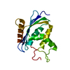
| ||||||||
|---|---|---|---|---|---|---|---|---|---|
| 1 |
| ||||||||
| Unit cell |
|
-Components
| #1: Protein | Mass: 17646.057 Da / Num. of mol.: 1 / Mutation: D134H Source method: isolated from a genetically manipulated source Source: (gene. exp.) |
|---|---|
| #2: Water | ChemComp-HOH /  Mass: 18.015 Da / Num. of mol.: 177 / Source method: isolated from a natural source / Formula: H2O Mass: 18.015 Da / Num. of mol.: 177 / Source method: isolated from a natural source / Formula: H2O |
-Experimental details
-Experiment
| Experiment | Method: X-RAY DIFFRACTION |
|---|
- Sample preparation
| Crystal | Density Matthews: 1.93 Å3/Da / Density % sol: 36.5 % | ||||||||||||||||||||
|---|---|---|---|---|---|---|---|---|---|---|---|---|---|---|---|---|---|---|---|---|---|
| Crystal grow | *PLUS Temperature: 20 ℃ / pH: 9 / Method: vapor diffusion, hanging drop / Details: macro-seeding | ||||||||||||||||||||
| Components of the solutions | *PLUS
|
-Data collection
| Diffraction source | Wavelength: 1.5418 |
|---|---|
| Detector | Type: ENRAF-NONIUS FAST / Detector: DIFFRACTOMETER / Date: May 13, 1993 |
| Radiation | Monochromatic (M) / Laue (L): M / Scattering type: x-ray |
| Radiation wavelength | Wavelength: 1.5418 Å / Relative weight: 1 |
| Reflection | Num. obs: 9295 / % possible obs: 81.9 % / Observed criterion σ(I): 0.5 / Redundancy: 3.07 % / Rmerge(I) obs: 0.077 |
| Reflection | *PLUS Highest resolution: 1.9 Å / Num. measured all: 28545 |
| Reflection shell | *PLUS Highest resolution: 1.9 Å / Lowest resolution: 1.93 Å / % possible obs: 68 % |
- Processing
| Software |
| ||||||||||||||||||||||||||||||||||||||||||||||||||||||||||||||||||||||||||||||||||||
|---|---|---|---|---|---|---|---|---|---|---|---|---|---|---|---|---|---|---|---|---|---|---|---|---|---|---|---|---|---|---|---|---|---|---|---|---|---|---|---|---|---|---|---|---|---|---|---|---|---|---|---|---|---|---|---|---|---|---|---|---|---|---|---|---|---|---|---|---|---|---|---|---|---|---|---|---|---|---|---|---|---|---|---|---|---|
| Refinement | Resolution: 1.9→6 Å / σ(F): 1 Details: IDEAL BOND LENGTHS AND ANGLES USED DURING REFINEMENT: HENDRICKSON AND KONNERT INITIAL REFINEMENTS WERE DONE WITH X-PLOR 3.1 BY BRUNGER.
| ||||||||||||||||||||||||||||||||||||||||||||||||||||||||||||||||||||||||||||||||||||
| Refinement step | Cycle: LAST / Resolution: 1.9→6 Å
| ||||||||||||||||||||||||||||||||||||||||||||||||||||||||||||||||||||||||||||||||||||
| Refine LS restraints |
| ||||||||||||||||||||||||||||||||||||||||||||||||||||||||||||||||||||||||||||||||||||
| Software | *PLUS Name: PROLSQ / Classification: refinement | ||||||||||||||||||||||||||||||||||||||||||||||||||||||||||||||||||||||||||||||||||||
| Refinement | *PLUS Rfactor obs: 0.197 | ||||||||||||||||||||||||||||||||||||||||||||||||||||||||||||||||||||||||||||||||||||
| Solvent computation | *PLUS | ||||||||||||||||||||||||||||||||||||||||||||||||||||||||||||||||||||||||||||||||||||
| Displacement parameters | *PLUS |
