 Movie
Movie Controller

[English] 日本語
 Yorodumi
- PDB-4jit: Crystal Structure of E. coli XGPRT in complex with (S)-3-(Guanin-... -
+ Open data

- Basic information
| Entry | Database: PDB / ID: 4jit | ||||||
|---|---|---|---|---|---|---|---|
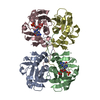
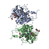
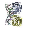
| Title | Crystal Structure of E. coli XGPRT in complex with (S)-3-(Guanin-9-yl)pyrrolidin-N-ylacetylphosphonic acid | ||||||
 Components Components | Xanthine phosphoribosyltransferase | ||||||
 Keywords Keywords | TRANSFERASE / XANTHINE-GUANINE PHOSPHORIBOSYLTRANSFERASE / PURINE SALVAGE / NUCLEOSIDE PHOSPHONATE / ANTIBACTERIAL | ||||||
| Function / homology | Rossmann fold - #2020 / Rossmann fold / 3-Layer(aba) Sandwich / Alpha Beta / Chem-3ZF / :  Function and homology information Function and homology information | ||||||
| Biological species |  | ||||||
| Method |  X-RAY DIFFRACTION / SYNCHROTRON / MOLECULAR REPLACEMENT / Resolution: 2.8 Å X-RAY DIFFRACTION / SYNCHROTRON / MOLECULAR REPLACEMENT / Resolution: 2.8 Å | ||||||
 Authors Authors | Keough, D.T. / Hockova, D. / Rejman, D. / Spacek, P. / Vrbkova, S. / Krecmerova, M. / Eng, W.S. / Jans, H. / West, N.P. / Naesens, L.M.J. ...Keough, D.T. / Hockova, D. / Rejman, D. / Spacek, P. / Vrbkova, S. / Krecmerova, M. / Eng, W.S. / Jans, H. / West, N.P. / Naesens, L.M.J. / de Jersey, J. / Guddat, L.W. | ||||||
 Citation Citation | Journal: J.Med.Chem. / Year: 2013 Title: Inhibition of the Escherichia coli 6-oxopurine phosphoribosyltransferases by nucleoside phosphonates: potential for new antibacterial agents. Authors: Keough, D.T. / Hockova, D. / Rejman, D. / Spacek, P. / Vrbkova, S. / Krecmerova, M. / Eng, W.S. / Jans, H. / West, N.P. / Naesens, L.M. / de Jersey, J. / Guddat, L.W. | ||||||
| History |
|
- Structure visualization
| Structure viewer | Molecule: MolmilJmol/JSmol |
|---|
- Downloads & links
-Download
| PDBx/mmCIF format | 4jit.cif.gz | 239 KB | Display | PDBx/mmCIF format |
|---|---|---|---|---|
| PDB format | pdb4jit.ent.gz | 196.4 KB | Display | PDB format |
| PDBx/mmJSON format | 4jit.json.gz | Tree view | PDBx/mmJSON format | |
| Others |  Other downloads Other downloads |
-Validation report
| Arichive directory | https://data.pdbj.org/pub/pdb/validation_reports/ji/4jitftp://data.pdbj.org/pub/pdb/validation_reports/ji/4jit | HTTPS FTP |
|---|
-Related structure data


-Links
 PDBj
PDBj- Assembly
| Deposited unit | 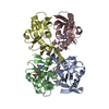
| ||||||||
|---|---|---|---|---|---|---|---|---|---|
| 1 |
| ||||||||
| Unit cell |
|
-Components
| #1: Protein | Mass: 16991.568 Da / Num. of mol.: 4 Source method: isolated from a genetically manipulated source Source: (gene. exp.) References: UniProt: H0Q6L9, xanthine phosphoribosyltransferase #2: Chemical |   Mass: 342.248 Da / Num. of mol.: 2 / Source method: obtained synthetically / Formula: C11H15N6O5P Mass: 342.248 Da / Num. of mol.: 2 / Source method: obtained synthetically / Formula: C11H15N6O5P#3: Water | ChemComp-HOH / |  Mass: 18.015 Da / Num. of mol.: 2 / Source method: isolated from a natural source / Formula: H2O Mass: 18.015 Da / Num. of mol.: 2 / Source method: isolated from a natural source / Formula: H2O |
|---|
-Experimental details
-Experiment
| Experiment | Method: X-RAY DIFFRACTION / Number of used crystals: 1 |
|---|
- Sample preparation
| Crystal | Density Matthews: 2.65 Å3/Da / Density % sol: 53.53 % |
|---|---|
| Crystal grow | Method: vapor diffusion, hanging drop / pH: 7 Details: 8% tacsimate, pH 7.0, 20% PEG 3350, VAPOR DIFFUSION, HANGING DROP |
-Data collection
| Diffraction | Mean temperature: 100 K |
|---|---|
| Diffraction source | Source: SYNCHROTRON / Site: Australian Synchrotron  / Beamline: MX2 / Wavelength: 0.95369 Å / Beamline: MX2 / Wavelength: 0.95369 Å |
| Detector | Type: ADSC QUANTUM 270 / Detector: CCD / Date: May 6, 2011 |
| Radiation | Monochromator: Si / Protocol: SINGLE WAVELENGTH / Monochromatic (M) / Laue (L): M / Scattering type: x-ray |
| Radiation wavelength | Wavelength: 0.95369 Å / Relative weight: 1 |
| Reflection | Resolution: 2.8→20 Å / Num. all: 18634 / Num. obs: 18634 / % possible obs: 100 % / Observed criterion σ(F): 0 / Observed criterion σ(I): 0 |
| Reflection shell | Resolution: 2.8→2.8756 Å / % possible all: 99.7 |
- Processing
| Software |
| ||||||||||||||||||||||||||||||||||||||||||||||||||||||||||||||||||||||||||||||||||||||||||||||||||
|---|---|---|---|---|---|---|---|---|---|---|---|---|---|---|---|---|---|---|---|---|---|---|---|---|---|---|---|---|---|---|---|---|---|---|---|---|---|---|---|---|---|---|---|---|---|---|---|---|---|---|---|---|---|---|---|---|---|---|---|---|---|---|---|---|---|---|---|---|---|---|---|---|---|---|---|---|---|---|---|---|---|---|---|---|---|---|---|---|---|---|---|---|---|---|---|---|---|---|---|
| Refinement | Method to determine structure: MOLECULAR REPLACEMENT / Resolution: 2.8→19.87 Å / SU ML: 0.31 / σ(F): 1.34 / Phase error: 29.25 / Stereochemistry target values: ML
| ||||||||||||||||||||||||||||||||||||||||||||||||||||||||||||||||||||||||||||||||||||||||||||||||||
| Solvent computation | Shrinkage radii: 0.9 Å / VDW probe radii: 1.11 Å / Solvent model: FLAT BULK SOLVENT MODEL | ||||||||||||||||||||||||||||||||||||||||||||||||||||||||||||||||||||||||||||||||||||||||||||||||||
| Refinement step | Cycle: LAST / Resolution: 2.8→19.87 Å
| ||||||||||||||||||||||||||||||||||||||||||||||||||||||||||||||||||||||||||||||||||||||||||||||||||
| Refine LS restraints |
| ||||||||||||||||||||||||||||||||||||||||||||||||||||||||||||||||||||||||||||||||||||||||||||||||||
| LS refinement shell |
| ||||||||||||||||||||||||||||||||||||||||||||||||||||||||||||||||||||||||||||||||||||||||||||||||||
| Refinement TLS params. | Method: refined / Origin x: -24.3803 Å / Origin y: 24.8837 Å / Origin z: -2.2408 Å
| ||||||||||||||||||||||||||||||||||||||||||||||||||||||||||||||||||||||||||||||||||||||||||||||||||
| Refinement TLS group |
|