 Movie
Movie Controller


+ Open data

- Basic information
| Entry | Database: PDB / ID: 4ivn | ||||||
|---|---|---|---|---|---|---|---|
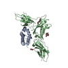
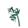
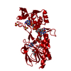
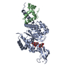
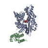
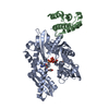
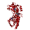
| Title | The Vibrio vulnificus NanR protein complexed with ManNAc-6P | ||||||
 Components Components | Transcriptional regulator Transcriptional regulation Transcriptional regulation | ||||||
 Keywords Keywords | TRANSCRIPTION REGULATOR / Isomerase fold / nan operon regulator for sialic acid catabolism | ||||||
| Function / homology |  Function and homology information Function and homology informationcarbohydrate derivative metabolic process / carbohydrate derivative binding / DNA-binding transcription repressor activity / protein-DNA complex / transcription cis-regulatory region binding / negative regulation of DNA-templated transcriptionSimilarity search - Function | ||||||
| Biological species |  Vibrio vulnificus (bacteria) Vibrio vulnificus (bacteria) | ||||||
| Method | X-RAY DIFFRACTION / SYNCHROTRON / MOLECULAR REPLACEMENT / Resolution: 1.9 Å | ||||||
 Authors Authors | Hwang, J. / Kim, M.H. | ||||||
 Citation Citation | Journal: Proc.Natl.Acad.Sci.USA / Year: 2013 Title: Structural insights into the regulation of sialic acid catabolism by the Vibrio vulnificus transcriptional repressor NanR Authors: Hwang, J. / Kim, B.S. / Jang, S.Y. / Lim, J.G. / You, D.J. / Jung, H.S. / Oh, T.K. / Lee, J.O. / Choi, S.H. / Kim, M.H. | ||||||
| History |
|
- Structure visualization
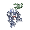
| Structure viewer | Molecule: MolmilJmol/JSmol |
|---|
- Downloads & links
-Download
| PDBx/mmCIF format | 4ivn.cif.gz | 212.9 KB | Display | PDBx/mmCIF format |
|---|---|---|---|---|
| PDB format | pdb4ivn.ent.gz | 171.8 KB | Display | PDB format |
| PDBx/mmJSON format | 4ivn.json.gz | Tree view | PDBx/mmJSON format | |
| Others |  Other downloads Other downloads |
-Validation report
| Arichive directory | https://data.pdbj.org/pub/pdb/validation_reports/iv/4ivnftp://data.pdbj.org/pub/pdb/validation_reports/iv/4ivn | HTTPS FTP |
|---|
-Related structure data
| Similar structure data |
|---|
-Links
 PDBj
PDBj- Assembly
| Deposited unit | 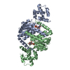
| ||||||||||||||||||
|---|---|---|---|---|---|---|---|---|---|---|---|---|---|---|---|---|---|---|---|
| 1 |
| ||||||||||||||||||
| Unit cell |
| ||||||||||||||||||
| Noncrystallographic symmetry (NCS) | NCS domain:
NCS domain segments: Component-ID: 0 / Ens-ID: 1 / Beg auth comp-ID: ASN / Beg label comp-ID: ASN / End auth comp-ID: ALA / End label comp-ID: ALA / Refine code: 0 / Auth seq-ID: 6 - 277 / Label seq-ID: 6 - 277
|
-Components
| #1: Protein | Transcriptional regulation / NanR Mass: 30341.633 Da / Num. of mol.: 2 Source method: isolated from a genetically manipulated source Source: (gene. exp.) Vibrio vulnificus (bacteria) / Strain: YJ016 / Production host: Escherichia coli (E. coli) / References: UniProt: Q7MD38#2: Sugar |   Type: D-saccharide, alpha linking / Mass: 301.188 Da / Num. of mol.: 2 Type: D-saccharide, alpha linking / Mass: 301.188 Da / Num. of mol.: 2Source method: isolated from a genetically manipulated source Formula: C8H16NO9P #3: Water | ChemComp-HOH / | Water Mass: 18.015 Da / Num. of mol.: 221 / Source method: isolated from a natural source / Formula: H2O Mass: 18.015 Da / Num. of mol.: 221 / Source method: isolated from a natural source / Formula: H2O |
|---|
-Experimental details
-Experiment
| Experiment | Method: X-RAY DIFFRACTION / Number of used crystals: 1 |
|---|
- Sample preparation
| Crystal | Density Matthews: 2.34 Å3/Da / Density % sol: 47.42 % |
|---|---|
| Crystal grow | Temperature: 294.15 K / Method: vapor diffusion, sitting drop / pH: 5 Details: 10% PEG 2000 MME, 0.1M ammonium sulfate, 0.3M sodium formate, 3% PGA-LM, 0.1M sodium acetate, pH 5.0, VAPOR DIFFUSION, SITTING DROP, temperature 294.15K |
-Data collection
| Diffraction | Mean temperature: 100.15 K |
|---|---|
| Diffraction source | Source: SYNCHROTRON / Site: Photon Factory  / Beamline: BL-17A / Wavelength: 1 Å / Beamline: BL-17A / Wavelength: 1 Å |
| Detector | Detector: CCD / Date: Jun 8, 2012 |
| Radiation | Protocol: SINGLE WAVELENGTH / Monochromatic (M) / Laue (L): M / Scattering type: x-ray |
| Radiation wavelength | Wavelength: 1 Å / Relative weight: 1 |
| Reflection | Resolution: 1.9→50 Å / Num. obs: 45033 / % possible obs: 99.9 % / Observed criterion σ(F): 2 / Observed criterion σ(I): 2 |
| Reflection shell | Resolution: 1.9→1.93 Å / % possible all: 100 |
- Processing
| Software | Name: REFMAC / Version: 5.6.0117 / Classification: refinement | |||||||||||||||||||||||||||||||||||||||||||||||||||||||||||||||||||||||||||||||||||||||||||||||||||||||||||||||||||||||||||||
|---|---|---|---|---|---|---|---|---|---|---|---|---|---|---|---|---|---|---|---|---|---|---|---|---|---|---|---|---|---|---|---|---|---|---|---|---|---|---|---|---|---|---|---|---|---|---|---|---|---|---|---|---|---|---|---|---|---|---|---|---|---|---|---|---|---|---|---|---|---|---|---|---|---|---|---|---|---|---|---|---|---|---|---|---|---|---|---|---|---|---|---|---|---|---|---|---|---|---|---|---|---|---|---|---|---|---|---|---|---|---|---|---|---|---|---|---|---|---|---|---|---|---|---|---|---|---|
| Refinement | Method to determine structure: MOLECULAR REPLACEMENT / Resolution: 1.9→30 Å / Cor.coef. Fo:Fc: 0.966 / Cor.coef. Fo:Fc free: 0.941 / SU B: 7.038 / SU ML: 0.106 / Cross valid method: THROUGHOUT / ESU R: 0.142 / ESU R Free: 0.144 / Stereochemistry target values: MAXIMUM LIKELIHOOD
| |||||||||||||||||||||||||||||||||||||||||||||||||||||||||||||||||||||||||||||||||||||||||||||||||||||||||||||||||||||||||||||
| Solvent computation | Ion probe radii: 0.8 Å / Shrinkage radii: 0.8 Å / VDW probe radii: 1.2 Å / Solvent model: MASK | |||||||||||||||||||||||||||||||||||||||||||||||||||||||||||||||||||||||||||||||||||||||||||||||||||||||||||||||||||||||||||||
| Displacement parameters | Biso mean: 42.466 Å2
| |||||||||||||||||||||||||||||||||||||||||||||||||||||||||||||||||||||||||||||||||||||||||||||||||||||||||||||||||||||||||||||
| Refinement step | Cycle: LAST / Resolution: 1.9→30 Å
| |||||||||||||||||||||||||||||||||||||||||||||||||||||||||||||||||||||||||||||||||||||||||||||||||||||||||||||||||||||||||||||
| Refine LS restraints |
| |||||||||||||||||||||||||||||||||||||||||||||||||||||||||||||||||||||||||||||||||||||||||||||||||||||||||||||||||||||||||||||
| Refine LS restraints NCS | Ens-ID: 1 / Number: 366 / Refine-ID: X-RAY DIFFRACTION / Type: interatomic distance / Rms dev position: 0.15 Å / Weight position: 0.05
| |||||||||||||||||||||||||||||||||||||||||||||||||||||||||||||||||||||||||||||||||||||||||||||||||||||||||||||||||||||||||||||
| LS refinement shell | Resolution: 1.9→1.949 Å / Total num. of bins used: 20
| |||||||||||||||||||||||||||||||||||||||||||||||||||||||||||||||||||||||||||||||||||||||||||||||||||||||||||||||||||||||||||||
| Refinement TLS params. | Method: refined / Refine-ID: X-RAY DIFFRACTION
| |||||||||||||||||||||||||||||||||||||||||||||||||||||||||||||||||||||||||||||||||||||||||||||||||||||||||||||||||||||||||||||
| Refinement TLS group |
|
