DNA double-strand break processing involved in repair via single-strand annealing / BRCA1-C complex / single-stranded DNA endodeoxyribonuclease activity / DNA strand resection involved in replication fork processing / homologous recombination / blastocyst hatching / Impaired BRCA2 binding to PALB2 / mitotic G2/M transition checkpoint / HDR through MMEJ (alt-NHEJ) / Homologous DNA Pairing and Strand Exchange ...DNA double-strand break processing involved in repair via single-strand annealing / BRCA1-C complex / single-stranded DNA endodeoxyribonuclease activity / DNA strand resection involved in replication fork processing / homologous recombination / blastocyst hatching / Impaired BRCA2 binding to PALB2 / mitotic G2/M transition checkpoint / HDR through MMEJ (alt-NHEJ) / Homologous DNA Pairing and Strand Exchange / Defective homologous recombination repair (HRR) due to BRCA1 loss of function / Defective HDR through Homologous Recombination Repair (HRR) due to PALB2 loss of BRCA1 binding function / Defective HDR through Homologous Recombination Repair (HRR) due to PALB2 loss of BRCA2/RAD51/RAD51C binding function / Resolution of D-loop Structures through Synthesis-Dependent Strand Annealing (SDSA) / Resolution of D-loop Structures through Holliday Junction Intermediates / HDR through Single Strand Annealing (SSA) / Impaired BRCA2 binding to RAD51 / Transcriptional Regulation by E2F6 / Presynaptic phase of homologous DNA pairing and strand exchange / transcription repressor complex / meiotic cell cycle / G1/S transition of mitotic cell cycle / double-strand break repair via homologous recombination / G2/M DNA damage checkpoint / HDR through Homologous Recombination (HRR) / Meiotic recombination / transcription corepressor activity / site of double-strand break / Processing of DNA double-strand break ends / Regulation of TP53 Activity through Phosphorylation / damaged DNA binding / Hydrolases; Acting on ester bonds / RNA polymerase II-specific DNA-binding transcription factor binding / cell division / nucleoplasm / metal ion binding / identical protein binding / nucleus Similarity search - Function
DNA endonuclease Ctp1, N-terminal / DNA endonuclease RBBP8-like / Tumour-suppressor protein CtIP N-terminal domain / DNA endonuclease activator Ctp1, C-terminal / DNA endonuclease activator SAE2/CtIP C-terminus Similarity search - Domain/homology
RBBP8 / CTBP-INTERACTING PROTEIN / CTIP / RETINOBLASTOMA-BINDING PROTEIN 8 / RBBP-8 / RETINOBLASTOMA- ...CTBP-INTERACTING PROTEIN / CTIP / RETINOBLASTOMA-BINDING PROTEIN 8 / RBBP-8 / RETINOBLASTOMA-INTERACTING PROTEIN AND MYOSIN-LIKE / RIM / SPORULATION IN THE ABSENCE OF SPO11 PROTEIN 2 HOMOLOG / SAE2
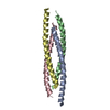
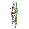
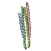
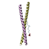
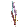
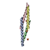
Mass: 4512.235 Da / Num. of mol.: 8 / Fragment: TETRAMERISATION DOMAIN, RESIDUES 18-52 Source method: isolated from a genetically manipulated source Source: (gene. exp.) HOMO SAPIENS (human) / Plasmid: PMAT11 / Production host: ESCHERICHIA COLI (E. coli) / Strain (production host): BL21(DE3) / Variant (production host): ROSETTA2 References: UniProt: Q99708, Hydrolases; Acting on ester bonds
Protocol: SINGLE WAVELENGTH / Monochromatic (M) / Laue (L): M / Scattering type: x-ray
Radiation wavelength
Wavelength: 1.0397 Å / Relative weight: 1
Reflection
Resolution: 1.9→28.98 Å / Num. obs: 20994 / % possible obs: 99.5 % / Observed criterion σ(I): 0 / Redundancy: 3.6 % / Biso Wilson estimate: 33.53 Å2 / Rmerge(I) obs: 0.06 / Net I/σ(I): 10.2
Reflection shell
Resolution: 1.9→1.94 Å / Redundancy: 3.6 % / Rmerge(I) obs: 0.92 / Mean I/σ(I) obs: 1.6 / % possible all: 99.5
-
Processing
Software
Name
Version
Classification
BUSTER
2.10.0
refinement
XDS
datareduction
XDS
datascaling
Aimless
datascaling
PHASER
phasing
Refinement
Method to determine structure: MOLECULAR REPLACEMENT / Resolution: 1.9→20.16 Å / Cor.coef. Fo:Fc: 0.9349 / Cor.coef. Fo:Fc free: 0.924 / SU R Cruickshank DPI: 0.492 / Cross valid method: THROUGHOUT / σ(F): 0 / SU R Blow DPI: 0.19 / SU Rfree Blow DPI: 0.162 / SU Rfree Cruickshank DPI: 0.164 Details: IDEAL-DIST CONTACT TERM CONTACT SETUP. ALL ATOMS HAVE CCP4 ATOM TYPE FROM LIBRARY.
Rfactor
Num. reflection
% reflection
Selection details
Rfree
0.2514
1079
5.15 %
RANDOM
Rwork
0.2122
-
-
-
obs
0.2144
20969
99.4 %
-
Displacement parameters
Biso mean: 49.38 Å2
Baniso -1
Baniso -2
Baniso -3
1-
-0.1078 Å2
0 Å2
0.4275 Å2
2-
-
-5.4139 Å2
0 Å2
3-
-
-
5.5217 Å2
Refinement step
Cycle: LAST / Resolution: 1.9→20.16 Å
Protein
Nucleic acid
Ligand
Solvent
Total
Num. atoms
2267
0
70
137
2474
Refine LS restraints
Refine-ID
Type
Dev ideal
Number
Restraint function
Weight
X-RAY DIFFRACTION
t_bond_d
0.01
4703
HARMONIC
2
X-RAY DIFFRACTION
t_angle_deg
0.88
8552
HARMONIC
2
X-RAY DIFFRACTION
t_dihedral_angle_d
1107
SINUSOIDAL
2
X-RAY DIFFRACTION
t_incorr_chiral_ct
X-RAY DIFFRACTION
t_pseud_angle
X-RAY DIFFRACTION
t_trig_c_planes
78
HARMONIC
2
X-RAY DIFFRACTION
t_gen_planes
623
HARMONIC
5
X-RAY DIFFRACTION
t_it
4703
HARMONIC
20
X-RAY DIFFRACTION
t_nbd
X-RAY DIFFRACTION
t_omega_torsion
2.35
X-RAY DIFFRACTION
t_other_torsion
14.2
X-RAY DIFFRACTION
t_improper_torsion
X-RAY DIFFRACTION
t_chiral_improper_torsion
279
SEMIHARMONIC
5
X-RAY DIFFRACTION
t_sum_occupancies
X-RAY DIFFRACTION
t_utility_distance
X-RAY DIFFRACTION
t_utility_angle
X-RAY DIFFRACTION
t_utility_torsion
X-RAY DIFFRACTION
t_ideal_dist_contact
4747
SEMIHARMONIC
4
LS refinement shell
Resolution: 1.9→1.99 Å / Total num. of bins used: 11
Rfactor
Num. reflection
% reflection
Rfree
0.2294
136
4.9 %
Rwork
0.2164
2642
-
all
0.217
2778
-
obs
-
-
99.4 %
Refinement TLS params.
Method: refined / Refine-ID: X-RAY DIFFRACTION
ID
L11 (°2)
L12 (°2)
L13 (°2)
L22 (°2)
L23 (°2)
L33 (°2)
S11 (Å °)
S12 (Å °)
S13 (Å °)
S21 (Å °)
S22 (Å °)
S23 (Å °)
S31 (Å °)
S32 (Å °)
S33 (Å °)
T11 (Å2)
T12 (Å2)
T13 (Å2)
T22 (Å2)
T23 (Å2)
T33 (Å2)
Origin x (Å)
Origin y (Å)
Origin z (Å)
1
1.7067
-0.3173
-0.4709
0.8468
-3.0063
13.8012
-0.0751
0.4117
-0.1381
-0.2985
0.1932
-0.2335
0.3318
-0.1157
-0.118
0.2053
-0.0748
0.0848
0.08
-0.0325
-0.2236
3.4807
-1.7288
9.7623
2
2.3703
0.3113
2.0236
2.4515
-0.1582
7.6394
-0.0575
-0.1392
0.0991
0.5208
-0.0601
-0.4378
0.0489
0.1926
0.1176
-0.0524
0.0318
-0.1298
-0.1508
-0.0527
-0.2177
5.0793
2.0162
36.9078
3
0.3807
0.2005
-1.3083
1.55
-1.5864
4.8291
-0.022
-0.0844
-0.0734
-0.0895
0.0374
0.0517
0.4498
-0.2257
-0.0154
-0.0693
-0.0292
-0.0333
0.1211
0.0077
-0.1857
-4.089
-5.4603
41.6799
4
0.9232
-0.2196
2.1618
0.6906
-1.0586
9.5247
0.04
-0.0652
-0.0229
-0.2471
0.0326
0.1167
-0.2366
-0.4973
-0.0727
0.0895
0.0779
-0.0749
0.0776
-0.001
-0.1379
-4.9876
4.8806
11.0018
5
1.6474
-0.6549
-0.9086
0.7689
0.2042
8.6547
-0.0217
-0.3968
-0.0015
0.0193
0.1747
-0.0077
-0.4601
0.4258
-0.153
0.0046
0.0335
-0.0634
0.0697
0.0056
-0.2688
22.6118
-16.1381
35.4284
6
0.9094
0.2972
-0.599
0.4446
0.0719
1.9523
0.0713
0.2117
-0.0411
-0.0842
0.0028
-0.1121
0.1387
0.2058
-0.074
0.1287
0.1529
-0.0984
0.1191
-0.0597
-0.2575
23.4812
-20.8514
11.8281
7
0.5544
0.301
-0.3566
0.8686
-0.931
6.8575
-0.1363
0.1916
-0.0542
-0.4709
0.1406
0.1061
-0.3138
-0.6478
-0.0042
0.2436
0.0312
-0.1349
0.119
-0.0483
-0.2233
14.3363
-13.4918
8.7485
8
0.4081
-0.187
0.2824
0.8057
-0.494
4.4101
0.0036
-0.0944
-0.1149
-0.2434
-0.0986
0.143
0.0797
-0.45
0.095
-0.0289
0.0304
-0.1182
0.0574
-0.0535
-0.1807
13.6081
-23.5621
33.8882
Refinement TLS group
ID
Refine-ID
Refine TLS-ID
Selection details
1
X-RAY DIFFRACTION
1
CHAINA
2
X-RAY DIFFRACTION
2
CHAINB
3
X-RAY DIFFRACTION
3
CHAINC
4
X-RAY DIFFRACTION
4
CHAIND
5
X-RAY DIFFRACTION
5
CHAINE
6
X-RAY DIFFRACTION
6
CHAINF
7
X-RAY DIFFRACTION
7
CHAING
8
X-RAY DIFFRACTION
8
CHAINH
+
About Yorodumi
-
News
-
Feb 9, 2022. New format data for meta-information of EMDB entries
New format data for meta-information of EMDB entries
Version 3 of the EMDB header file is now the official format.
The previous official version 1.9 will be removed from the archive.
In the structure databanks used in Yorodumi, some data are registered as the other names, "COVID-19 virus" and "2019-nCoV". Here are the details of the virus and the list of structure data.
Jan 31, 2019. EMDB accession codes are about to change! (news from PDBe EMDB page)
EMDB accession codes are about to change! (news from PDBe EMDB page)
The allocation of 4 digits for EMDB accession codes will soon come to an end. Whilst these codes will remain in use, new EMDB accession codes will include an additional digit and will expand incrementally as the available range of codes is exhausted. The current 4-digit format prefixed with “EMD-” (i.e. EMD-XXXX) will advance to a 5-digit format (i.e. EMD-XXXXX), and so on. It is currently estimated that the 4-digit codes will be depleted around Spring 2019, at which point the 5-digit format will come into force.
The EM Navigator/Yorodumi systems omit the EMD- prefix.
Related info.:Q: What is EMD? / ID/Accession-code notation in Yorodumi/EM Navigator
Yorodumi is a browser for structure data from EMDB, PDB, SASBDB, etc.
This page is also the successor to EM Navigator detail page, and also detail information page/front-end page for Omokage search.
The word "yorodu" (or yorozu) is an old Japanese word meaning "ten thousand". "mi" (miru) is to see.
Related info.:EMDB / PDB / SASBDB / Comparison of 3 databanks / Yorodumi Search / Aug 31, 2016. New EM Navigator & Yorodumi / Yorodumi Papers / Jmol/JSmol / Function and homology information / Changes in new EM Navigator and Yorodumi
 Movie
Movie Controller


 Open data
 Basic information
 Components
 Keywords
 Function and homology information
 HOMO SAPIENS (human)
 X-RAY DIFFRACTION /
  Authors
 Citation
 Structure visualization
 Downloads & links
 Other downloads
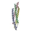


 PDBj
PDBj


 Assembly
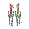


 Mass: 96.063 Da / Num. of mol.: 14 / Source method: obtained synthetically / Formula: SO4
 Mass: 18.015 Da / Num. of mol.: 137 / Source method: isolated from a natural source / Formula: H2O
 Sample preparation
 / Beamline: PROXIMA 1 / Wavelength: 1.0397
  Processing