 Movie
Movie Controller

[English] 日本語
 Yorodumi
- PDB-4baf: Hen egg-white lysozyme structure in complex with the europium tri... -
+ Open data

- Basic information
| Entry | Database: PDB / ID: 4baf | ||||||
|---|---|---|---|---|---|---|---|
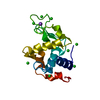
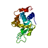
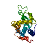
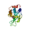
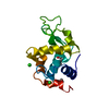
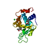
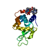
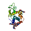
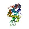
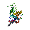
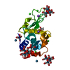
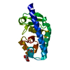
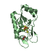
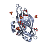
| Title | Hen egg-white lysozyme structure in complex with the europium tris- hydroxyethyltriazoledipicolinate complex at 1.51 A resolution. | ||||||
 Components Components | LYSOZYME C | ||||||
 Keywords Keywords | HYDROLASE / CLICK-CHEMISTRY / ANOMALOUS SCATTERING / DE NOVO PHASING / LANTHANIDE COMPLEX / DIPICOLINATE | ||||||
| Function / homology |  Function and homology information Function and homology informationLactose synthesis / Antimicrobial peptides / Neutrophil degranulation / beta-N-acetylglucosaminidase activity / cell wall macromolecule catabolic process / lysozyme / lysozyme activity / killing of cells of another organism / defense response to Gram-negative bacterium / defense response to bacterium ...Lactose synthesis / Antimicrobial peptides / Neutrophil degranulation / beta-N-acetylglucosaminidase activity / cell wall macromolecule catabolic process / lysozyme / lysozyme activity / killing of cells of another organism / defense response to Gram-negative bacterium / defense response to bacterium / defense response to Gram-positive bacterium / endoplasmic reticulum / extracellular space / identical protein binding / cytoplasm Similarity search - Function | ||||||
| Biological species |  | ||||||
| Method |  X-RAY DIFFRACTION / SYNCHROTRON / MAD / Resolution: 1.507 Å X-RAY DIFFRACTION / SYNCHROTRON / MAD / Resolution: 1.507 Å | ||||||
 Authors Authors | Talon, R. / Kahn, R. / Gautier, A. / Nauton, L. / Girard, E. | ||||||
 Citation Citation | Journal: Chem.Commun.(Camb.) / Year: 2012 Title: Clicked Europium Dipicolinate Complexes for Protein X-Ray Structure Determination. Authors: Talon, R. / Nauton, L. / Canet, J.-L. / Kahn, R. / Girard, E. / Gautier, A. #1: Journal: Angew.Chem.Int.Ed.Engl. / Year: 2008Title: Protein Crystallography Through Supramolecular Interactions between a Lanthanide Complex and Arginine. Authors: Pompidor, G. / D'Aleo, A. / Vicat, J. / Toupet, L. / Giraud, N. / Kahn, R. / Maury, O. #2: Journal: Acta Crystallogr.,Sect.D / Year: 2010Title: A Dipicolinate Lanthanide Complex for Solving Protein Structures Using Anomalous Diffraction. Authors: Pompidor, G. / Maury, O. / Vicat, J. / Kahn, R. #3: Journal: Acta Crystallogr.,Sect.D / Year: 2002Title: Gd-Hpdo3A, a Complex to Obtain High-Phasing-Power Heavy-Atom Derivatives for Sad and MAD Experiments: Results with Tetragonal Hen Egg-White Lysozyme. Authors: Girard, E. / Chantalat, L. / Vicat, J. / Kahn, R. | ||||||
| History |
|
- Structure visualization
| Structure viewer | Molecule: MolmilJmol/JSmol |
|---|
- Downloads & links
-Download
| PDBx/mmCIF format | 4baf.cif.gz | 74.6 KB | Display | PDBx/mmCIF format |
|---|---|---|---|---|
| PDB format | pdb4baf.ent.gz | 56.1 KB | Display | PDB format |
| PDBx/mmJSON format | 4baf.json.gz | Tree view | PDBx/mmJSON format | |
| Others |  Other downloads Other downloads |
-Validation report
| Arichive directory | https://data.pdbj.org/pub/pdb/validation_reports/ba/4bafftp://data.pdbj.org/pub/pdb/validation_reports/ba/4baf | HTTPS FTP |
|---|
-Related structure data


-Links
 PDBj
PDBj


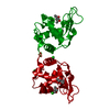
- Assembly
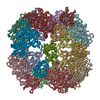
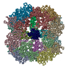
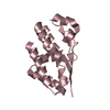
| Deposited unit | 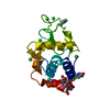
| ||||||||||||
|---|---|---|---|---|---|---|---|---|---|---|---|---|---|
| 1 |
| ||||||||||||
| Unit cell |
| ||||||||||||
| Components on special symmetry positions |
|
-Components
-Protein , 1 types, 1 molecules A
| #1: Protein | Mass: 14331.160 Da / Num. of mol.: 1 / Source method: isolated from a natural source Details: ROCHE APPLIED SCIENCE POWDER, CATALOG NUMBER 10837059001 Source: (natural) |
|---|
-Non-polymers , 6 types, 164 molecules 


| #2: Chemical | ChemComp-EU3 /  Mass: 151.964 Da / Num. of mol.: 1 / Source method: obtained synthetically / Formula: Eu Mass: 151.964 Da / Num. of mol.: 1 / Source method: obtained synthetically / Formula: Eu | ||||||||
|---|---|---|---|---|---|---|---|---|---|
| #3: Chemical |  Mass: 280.237 Da / Num. of mol.: 3 / Source method: obtained synthetically / Formula: C11H12N4O5 Mass: 280.237 Da / Num. of mol.: 3 / Source method: obtained synthetically / Formula: C11H12N4O5#4: Chemical | ChemComp-ACT / |  Mass: 59.044 Da / Num. of mol.: 1 / Source method: obtained synthetically / Formula: C2H3O2 Mass: 59.044 Da / Num. of mol.: 1 / Source method: obtained synthetically / Formula: C2H3O2#5: Chemical | ChemComp-CL /  Mass: 35.453 Da / Num. of mol.: 9 / Source method: obtained synthetically / Formula: Cl Mass: 35.453 Da / Num. of mol.: 9 / Source method: obtained synthetically / Formula: Cl#6: Chemical |  Mass: 22.990 Da / Num. of mol.: 2 / Source method: obtained synthetically / Formula: Na Mass: 22.990 Da / Num. of mol.: 2 / Source method: obtained synthetically / Formula: Na#7: Water | ChemComp-HOH / | Mass: 18.015 Da / Num. of mol.: 148 / Source method: isolated from a natural source / Formula: H2O |
-Details
| Has protein modification | Y |
|---|---|
| Sequence details | THE SEQUENCE PROVIDED CORRESPOND |
-Experimental details
-Experiment
| Experiment | Method: X-RAY DIFFRACTION / Number of used crystals: 1 |
|---|
- Sample preparation
| Crystal | Density Matthews: 1.68 Å3/Da / Density % sol: 27 % Description: MAD DATA AT TWO WAVELENGTHS WERE COLLECTED. THE FIRST DATA SET AT THE EUROPIUM LIII EDGE AND THE SECOND ONE AT THE SELENIUM K EDGE |
|---|---|
| Crystal grow | Temperature: 293 K / pH: 4.6 Details: 0.1 M SODIUM ACETATE PH 4.6, 0.4-1.5 M SODIUM CHLORIDE, 0.00046 M PROTEIN, 0.001.4 M LANTHANIDE COMPLEX, 293 K, 3-7 DAYS |
-Data collection
| Diffraction | Mean temperature: 100 K |
|---|---|
| Diffraction source | Source: SYNCHROTRON / Site: ESRF  / Beamline: BM30A / Wavelength: 0.979 / Beamline: BM30A / Wavelength: 0.979 |
| Detector | Type: ADSC CCD / Detector: CCD / Date: Nov 6, 2009 / Details: MIRRORS |
| Radiation | Monochromator: EITHER SI111 OR SI311 CRYSTALS / Protocol: MAD / Monochromatic (M) / Laue (L): M / Scattering type: x-ray |
| Radiation wavelength | Wavelength: 0.979 Å / Relative weight: 1 |
| Reflection | Resolution: 1.51→38.88 Å / Num. obs: 18899 / % possible obs: 99.7 % / Observed criterion σ(I): 3 / Redundancy: 13.6 % / Biso Wilson estimate: 13.89 Å2 / Rmerge(I) obs: 0.05 / Net I/σ(I): 10.3 |
| Reflection shell | Resolution: 1.51→1.59 Å / Redundancy: 12.7 % / Rmerge(I) obs: 0.32 / Mean I/σ(I) obs: 2.4 / % possible all: 98 |
- Processing
| Software |
| |||||||||||||||||||||||||||||||||||||||||||||||||||||||||||||||||||||||||||||||||||||||||||||||||||||||||||||||||||||||||||||||||||||||||||||||||||||||||||||||||||||||||||||||
|---|---|---|---|---|---|---|---|---|---|---|---|---|---|---|---|---|---|---|---|---|---|---|---|---|---|---|---|---|---|---|---|---|---|---|---|---|---|---|---|---|---|---|---|---|---|---|---|---|---|---|---|---|---|---|---|---|---|---|---|---|---|---|---|---|---|---|---|---|---|---|---|---|---|---|---|---|---|---|---|---|---|---|---|---|---|---|---|---|---|---|---|---|---|---|---|---|---|---|---|---|---|---|---|---|---|---|---|---|---|---|---|---|---|---|---|---|---|---|---|---|---|---|---|---|---|---|---|---|---|---|---|---|---|---|---|---|---|---|---|---|---|---|---|---|---|---|---|---|---|---|---|---|---|---|---|---|---|---|---|---|---|---|---|---|---|---|---|---|---|---|---|---|---|---|---|---|
| Refinement | Method to determine structure: MAD Starting model: NONE Resolution: 1.507→31.203 Å / SU ML: 0.29 / σ(F): 1.38 / Phase error: 13.38 / Stereochemistry target values: ML Details: LIGAND OCCUPANCIES WERE FIXED ACCORDING TO THE ANTHANIDE ION OCCUPANCY
| |||||||||||||||||||||||||||||||||||||||||||||||||||||||||||||||||||||||||||||||||||||||||||||||||||||||||||||||||||||||||||||||||||||||||||||||||||||||||||||||||||||||||||||||
| Solvent computation | Shrinkage radii: 0.83 Å / VDW probe radii: 1.1 Å / Solvent model: FLAT BULK SOLVENT MODEL / Bsol: 52.575 Å2 / ksol: 0.449 e/Å3 | |||||||||||||||||||||||||||||||||||||||||||||||||||||||||||||||||||||||||||||||||||||||||||||||||||||||||||||||||||||||||||||||||||||||||||||||||||||||||||||||||||||||||||||||
| Displacement parameters | Biso mean: 15.87 Å2
| |||||||||||||||||||||||||||||||||||||||||||||||||||||||||||||||||||||||||||||||||||||||||||||||||||||||||||||||||||||||||||||||||||||||||||||||||||||||||||||||||||||||||||||||
| Refinement step | Cycle: LAST / Resolution: 1.507→31.203 Å
| |||||||||||||||||||||||||||||||||||||||||||||||||||||||||||||||||||||||||||||||||||||||||||||||||||||||||||||||||||||||||||||||||||||||||||||||||||||||||||||||||||||||||||||||
| Refine LS restraints |
| |||||||||||||||||||||||||||||||||||||||||||||||||||||||||||||||||||||||||||||||||||||||||||||||||||||||||||||||||||||||||||||||||||||||||||||||||||||||||||||||||||||||||||||||
| LS refinement shell |
| |||||||||||||||||||||||||||||||||||||||||||||||||||||||||||||||||||||||||||||||||||||||||||||||||||||||||||||||||||||||||||||||||||||||||||||||||||||||||||||||||||||||||||||||
| Refinement TLS params. | Method: refined / Refine-ID: X-RAY DIFFRACTION
| |||||||||||||||||||||||||||||||||||||||||||||||||||||||||||||||||||||||||||||||||||||||||||||||||||||||||||||||||||||||||||||||||||||||||||||||||||||||||||||||||||||||||||||||
| Refinement TLS group |
|
