 Movie
Movie Controller


+ Open data

- Basic information
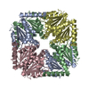
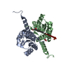
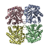
| Entry | Database: PDB / ID: 4ay4 | ||||||
|---|---|---|---|---|---|---|---|
| Title | crystal structure of Bacillus anthracis PurE | ||||||
 Components Components | N5-CARBOXYAMINOIMIDAZOLE RIBONUCLEOTIDE MUTASE | ||||||
 Keywords Keywords |  LYASE / ISOMERASE LYASE / ISOMERASE | ||||||
| Function / homology |  Function and homology information5-(carboxyamino)imidazole ribonucleotide mutase / 5-(carboxyamino)imidazole ribonucleotide mutase activity / 'de novo' IMP biosynthetic process / lyase activity Function and homology information5-(carboxyamino)imidazole ribonucleotide mutase / 5-(carboxyamino)imidazole ribonucleotide mutase activity / 'de novo' IMP biosynthetic process / lyase activitySimilarity search - Function | ||||||
| Biological species |  BACILLUS ANTHRACIS (anthrax bacterium) BACILLUS ANTHRACIS (anthrax bacterium) | ||||||
| Method | X-RAY DIFFRACTION / SYNCHROTRON / MOLECULAR REPLACEMENT / Resolution: 2 Å | ||||||
 Authors Authors | Oliete, R. / Pous, J. / Rodriguez-Puente, S. / Abad-Zapatero, C. / Guasch, A. | ||||||
 Citation Citation | Journal: Acta Crystallogr.,Sect.D / Year: 2013 Title: Elastic and Inelastic Diffraction Changes Upon Variation of the Relative Humidity Environment of Pure Crystals Authors: Oliete, R. / Pous, J. / Rodriguez-Puente, S. / Abad-Zapatero, C. / Guasch, A. | ||||||
| History |
|
- Structure visualization
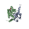
| Structure viewer | Molecule: MolmilJmol/JSmol |
|---|
- Downloads & links
-Download
| PDBx/mmCIF format | 4ay4.cif.gz | 248.2 KB | Display | PDBx/mmCIF format |
|---|---|---|---|---|
| PDB format | pdb4ay4.ent.gz | 201.3 KB | Display | PDB format |
| PDBx/mmJSON format | 4ay4.json.gz | Tree view | PDBx/mmJSON format | |
| Others |  Other downloads Other downloads |
-Validation report
| Arichive directory | https://data.pdbj.org/pub/pdb/validation_reports/ay/4ay4ftp://data.pdbj.org/pub/pdb/validation_reports/ay/4ay4 | HTTPS FTP |
|---|
-Related structure data
| Related structure data |  4ay3C  4b4kC 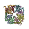 1xmpS S: Starting model for refinement C: citing same article ( |
|---|---|
| Similar structure data |

-Links
 PDBj
PDBj- Assembly
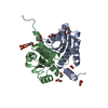
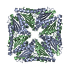
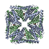
| Deposited unit | 
| ||||||||||||||||||||||||||||||||||||||||||||||||||||||||||||||||||||||||||||||||||||||||||||||||||||||||
|---|---|---|---|---|---|---|---|---|---|---|---|---|---|---|---|---|---|---|---|---|---|---|---|---|---|---|---|---|---|---|---|---|---|---|---|---|---|---|---|---|---|---|---|---|---|---|---|---|---|---|---|---|---|---|---|---|---|---|---|---|---|---|---|---|---|---|---|---|---|---|---|---|---|---|---|---|---|---|---|---|---|---|---|---|---|---|---|---|---|---|---|---|---|---|---|---|---|---|---|---|---|---|---|---|---|
| 1 | 
| ||||||||||||||||||||||||||||||||||||||||||||||||||||||||||||||||||||||||||||||||||||||||||||||||||||||||
| Unit cell |
| ||||||||||||||||||||||||||||||||||||||||||||||||||||||||||||||||||||||||||||||||||||||||||||||||||||||||
| Noncrystallographic symmetry (NCS) | NCS domain:
NCS domain segments: Beg auth comp-ID: LYS / Beg label comp-ID: LYS / Refine code: 1
NCS ensembles :
NCS oper:
|
-Components
| #1: Protein | Mass: 19232.014 Da / Num. of mol.: 4 Source method: isolated from a genetically manipulated source Source: (gene. exp.) BACILLUS ANTHRACIS (anthrax bacterium) / Production host: ESCHERICHIA COLI (E. coli) / Strain (production host): BL21(DE3)References: UniProt: Q81ZH8, UniProt: A0A6L7HA71*PLUS, phosphoribosylaminoimidazole carboxylase, 5-(carboxyamino)imidazole ribonucleotide mutase#2: Chemical | ChemComp-ACT / Acetate  Mass: 59.044 Da / Num. of mol.: 5 / Source method: obtained synthetically / Formula: C2H3O2 Mass: 59.044 Da / Num. of mol.: 5 / Source method: obtained synthetically / Formula: C2H3O2#3: Water | ChemComp-HOH / | Water Mass: 18.015 Da / Num. of mol.: 365 / Source method: isolated from a natural source / Formula: H2O Mass: 18.015 Da / Num. of mol.: 365 / Source method: isolated from a natural source / Formula: H2O |
|---|
-Experimental details
-Experiment
| Experiment | Method: X-RAY DIFFRACTION / Number of used crystals: 1 |
|---|
- Sample preparation
| Crystal | Density Matthews: 1.92 Å3/Da / Density % sol: 35.9 % / Description: NONE |
|---|---|
| Crystal grow | pH: 8.5 Details: PROTEIN WAS CRYSTALLIZED FROM 0.1M TRIS PH8.5, 0.3M SODIUM ACETATE, 15% PEG 4K |
-Data collection
| Diffraction | Mean temperature: 100 K |
|---|---|
| Diffraction source | Source: SYNCHROTRON / Site: ESRF  / Beamline: ID14-2 / Wavelength: 0.933 / Beamline: ID14-2 / Wavelength: 0.933 |
| Detector | Type: ADSC CCD / Detector: CCD / Date: Dec 12, 2008 / Details: TOROIDAL MIRRORS |
| Radiation | Monochromator: DIAMOND (111), GE(220) / Protocol: SINGLE WAVELENGTH / Monochromatic (M) / Laue (L): M / Scattering type: x-ray |
| Radiation wavelength | Wavelength: 0.933 Å / Relative weight: 1 |
| Reflection | Resolution: 2→50 Å / Num. obs: 42925 / % possible obs: 96.6 % / Observed criterion σ(I): -3 / Redundancy: 8.1 % / Rmerge(I) obs: 0.07 / Net I/σ(I): 32.66 |
| Reflection shell | Resolution: 2→2.07 Å / Redundancy: 6 % / Rmerge(I) obs: 0.29 / Mean I/σ(I) obs: 5.82 / % possible all: 94.1 |
- Processing
| Software |
| ||||||||||||||||||||||||||||||||||||||||||||||||||||||||||||||||||||||||||||||||||||||||||||||||||||||||||||||||||||||||||||||||||||||||||||||||||||||||||||||||||||||||||||||||||||||
|---|---|---|---|---|---|---|---|---|---|---|---|---|---|---|---|---|---|---|---|---|---|---|---|---|---|---|---|---|---|---|---|---|---|---|---|---|---|---|---|---|---|---|---|---|---|---|---|---|---|---|---|---|---|---|---|---|---|---|---|---|---|---|---|---|---|---|---|---|---|---|---|---|---|---|---|---|---|---|---|---|---|---|---|---|---|---|---|---|---|---|---|---|---|---|---|---|---|---|---|---|---|---|---|---|---|---|---|---|---|---|---|---|---|---|---|---|---|---|---|---|---|---|---|---|---|---|---|---|---|---|---|---|---|---|---|---|---|---|---|---|---|---|---|---|---|---|---|---|---|---|---|---|---|---|---|---|---|---|---|---|---|---|---|---|---|---|---|---|---|---|---|---|---|---|---|---|---|---|---|---|---|---|---|
| Refinement | Method to determine structure: MOLECULAR REPLACEMENT Starting model: PDB ENTRY 1XMP Resolution: 2→43.93 Å / Cor.coef. Fo:Fc: 0.967 / Cor.coef. Fo:Fc free: 0.953 / SU B: 5.778 / SU ML: 0.083 / Cross valid method: THROUGHOUT / ESU R: 0.161 / ESU R Free: 0.135 / Stereochemistry target values: MAXIMUM LIKELIHOOD Details: HYDROGENS HAVE BEEN ADDED IN THE RIDING POSITIONS. U VALUES WITH TLS ADDED.
| ||||||||||||||||||||||||||||||||||||||||||||||||||||||||||||||||||||||||||||||||||||||||||||||||||||||||||||||||||||||||||||||||||||||||||||||||||||||||||||||||||||||||||||||||||||||
| Solvent computation | Ion probe radii: 0.8 Å / Shrinkage radii: 0.8 Å / VDW probe radii: 1.2 Å / Solvent model: BABINET MODEL WITH MASK | ||||||||||||||||||||||||||||||||||||||||||||||||||||||||||||||||||||||||||||||||||||||||||||||||||||||||||||||||||||||||||||||||||||||||||||||||||||||||||||||||||||||||||||||||||||||
| Displacement parameters | Biso mean: 25.877 Å2
| ||||||||||||||||||||||||||||||||||||||||||||||||||||||||||||||||||||||||||||||||||||||||||||||||||||||||||||||||||||||||||||||||||||||||||||||||||||||||||||||||||||||||||||||||||||||
| Refinement step | Cycle: LAST / Resolution: 2→43.93 Å
| ||||||||||||||||||||||||||||||||||||||||||||||||||||||||||||||||||||||||||||||||||||||||||||||||||||||||||||||||||||||||||||||||||||||||||||||||||||||||||||||||||||||||||||||||||||||
| Refine LS restraints |
|