Class I PurE / PurE, prokaryotic type / PurE domain / AIR carboxylase / AIR carboxylase / Rossmann fold - #1970 / Rossmann fold / 3-Layer(aba) Sandwich / Alpha Beta Similarity search - Domain/homology
Resolution: 2.5→47.11 Å / Cor.coef. Fo:Fc: 0.946 / Cor.coef. Fo:Fc free: 0.919 / SU B: 18.26 / SU ML: 0.206 / Cross valid method: THROUGHOUT / ESU R Free: 0.295 / Stereochemistry target values: MAXIMUM LIKELIHOOD Details: HYDROGENS HAVE BEEN ADDED IN THE RIDING POSITIONS. U VALUES WITH TLS ADDED
Rfactor
Num. reflection
% reflection
Selection details
Rfree
0.21712
2878
5.1 %
RANDOM
Rwork
0.18013
-
-
-
obs
0.18209
53163
93.07 %
-
Solvent computation
Ion probe radii: 0.8 Å / Shrinkage radii: 0.8 Å / VDW probe radii: 1.2 Å / Solvent model: BABINET MODEL WITH MASK
 Movie
Movie Controller


 Open data
 Basic information
 Components
 Keywords
 Function and homology information
 X-RAY DIFFRACTION /
  Authors
 Citation
 Structure visualization
 Downloads & links
 Other downloads


 PDBj
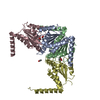
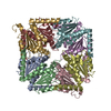
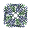
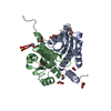
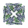
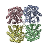
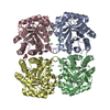
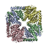
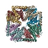
PDBj Assembly
 Mass: 18.015 Da / Num. of mol.: 483 / Source method: isolated from a natural source / Formula: H2O
 Sample preparation
 / Beamline: ID14-1 / Wavelength: 0.9334
  Processing