 Movie
Movie Controller

[English] 日本語
 Yorodumi
- PDB-4aim: Crystal structure of C. crescentus PNPase bound to RNase E recogn... -
+ Open data

- Basic information
| Entry | Database: PDB / ID: 4aim | ||||||
|---|---|---|---|---|---|---|---|
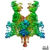
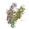
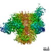
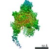
| Title | Crystal structure of C. crescentus PNPase bound to RNase E recognition peptide | ||||||
 Components Components |
| ||||||
 Keywords Keywords | TRANSFERASE/PEPTIDE / TRANSFERASE-PEPTIDE COMPLEX / KH DOMAIN / S1 DOMAIN / GWW PEPTIDE | ||||||
| Function / homology |  Function and homology information Function and homology informationribonuclease E / polyribonucleotide nucleotidyltransferase / polyribonucleotide nucleotidyltransferase activity / ribonuclease E activity / tRNA processing / mRNA catabolic process / RNA processing / RNA endonuclease activity / cytoplasmic side of plasma membrane / rRNA processing ...ribonuclease E / polyribonucleotide nucleotidyltransferase / polyribonucleotide nucleotidyltransferase activity / ribonuclease E activity / tRNA processing / mRNA catabolic process / RNA processing / RNA endonuclease activity / cytoplasmic side of plasma membrane / rRNA processing / 3'-5'-RNA exonuclease activity / tRNA binding / rRNA binding / magnesium ion binding / RNA binding / zinc ion binding / cytoplasm / cytosol Similarity search - Function | ||||||
| Biological species |  CAULOBACTER VIBRIOIDES (bacteria) CAULOBACTER VIBRIOIDES (bacteria) | ||||||
| Method |  X-RAY DIFFRACTION / SYNCHROTRON / MOLECULAR REPLACEMENT / Resolution: 3.3 Å X-RAY DIFFRACTION / SYNCHROTRON / MOLECULAR REPLACEMENT / Resolution: 3.3 Å | ||||||
 Authors Authors | Hardwick, S.W. / Gubbey, T. / Hug, I. / Jenal, U. / Luisi, B.F. | ||||||
 Citation Citation | Journal: Open Biol. / Year: 2012 Title: Crystal Structure of Caulobacter Crescentus Polynucleotide Phosphorylase Reveals a Mechanism of RNA Substrate Channelling and RNA Degradosome Assembly. Authors: Hardwick, S.W. / Gubbey, T. / Hug, I. / Jenal, U. / Luisi, B.F. | ||||||
| History |
|
- Structure visualization
| Structure viewer | Molecule: MolmilJmol/JSmol |
|---|
- Downloads & links
-Download
| PDBx/mmCIF format | 4aim.cif.gz | 157.6 KB | Display | PDBx/mmCIF format |
|---|---|---|---|---|
| PDB format | pdb4aim.ent.gz | 119.5 KB | Display | PDB format |
| PDBx/mmJSON format | 4aim.json.gz | Tree view | PDBx/mmJSON format | |
| Others |  Other downloads Other downloads |
-Validation report
| Summary document | 4aim_validation.pdf.gz | 450.7 KB | Display | wwPDB validaton report |
|---|---|---|---|---|
| Full document | 4aim_full_validation.pdf.gz | 487.5 KB | Display | |
| Data in XML | 4aim_validation.xml.gz | 30.8 KB | Display | |
| Data in CIF | 4aim_validation.cif.gz | 41.1 KB | Display | |
| Arichive directory | https://data.pdbj.org/pub/pdb/validation_reports/ai/4aimftp://data.pdbj.org/pub/pdb/validation_reports/ai/4aim | HTTPS FTP |
-Related structure data
| Related structure data |  4aidC  4am3C  3gmeS C: citing same article ( S: Starting model for refinement |
|---|---|
| Similar structure data |

-Links
 PDBj
PDBj


- Assembly
| Deposited unit | 
| ||||||||
|---|---|---|---|---|---|---|---|---|---|
| 1 | 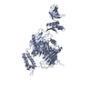
| ||||||||
| Unit cell |
|
-Components
| #1: Protein | Mass: 78145.383 Da / Num. of mol.: 1 Source method: isolated from a genetically manipulated source Source: (gene. exp.) CAULOBACTER VIBRIOIDES (bacteria) / Strain: CB15 / Production host: References: UniProt: Q9AC32, polyribonucleotide nucleotidyltransferase |
|---|---|
| #2: Protein/peptide | Mass: 1798.081 Da / Num. of mol.: 1 / Fragment: PNPASE BINDING PEPTIDE - GWW, RESIDUES 885-898 / Source method: obtained synthetically / Source: (synth.) CAULOBACTER VIBRIOIDES (bacteria) / References: UniProt: Q9A749 |
| #3: Chemical | ChemComp-PO4 /   Mass: 94.971 Da / Num. of mol.: 1 / Source method: obtained synthetically / Formula: PO4 Mass: 94.971 Da / Num. of mol.: 1 / Source method: obtained synthetically / Formula: PO4 |
| #4: Water | ChemComp-HOH /  Mass: 18.015 Da / Num. of mol.: 2 / Source method: isolated from a natural source / Formula: H2O Mass: 18.015 Da / Num. of mol.: 2 / Source method: isolated from a natural source / Formula: H2O |
-Experimental details
-Experiment
| Experiment | Method: X-RAY DIFFRACTION / Number of used crystals: 1 |
|---|
- Sample preparation
| Crystal | Density Matthews: 3.28 Å3/Da / Density % sol: 62.52 % / Description: NONE |
|---|---|
| Crystal grow | Details: 17 % WT/V PEG 3000, 0.1 M TRI-SODIUM CITRATE |
-Data collection
| Diffraction | Mean temperature: 100 K |
|---|---|
| Diffraction source | Source: SYNCHROTRON / Site: Diamond  / Beamline: I24 / Wavelength: 0.9778 / Beamline: I24 / Wavelength: 0.9778 |
| Detector | Type: MARRESEARCH MX-300 / Detector: CCD / Date: Oct 14, 2010 |
| Radiation | Protocol: SINGLE WAVELENGTH / Monochromatic (M) / Laue (L): M / Scattering type: x-ray |
| Radiation wavelength | Wavelength: 0.9778 Å / Relative weight: 1 |
| Reflection | Resolution: 3.3→40 Å / Num. obs: 15391 / % possible obs: 99.3 % / Observed criterion σ(I): 2 / Redundancy: 4.7 % / Rmerge(I) obs: 0.14 / Net I/σ(I): 7.8 |
| Reflection shell | Resolution: 3.3→3.48 Å / Redundancy: 4.6 % / Rmerge(I) obs: 0.65 / Mean I/σ(I) obs: 2.4 / % possible all: 99.2 |
- Processing
| Software |
| ||||||||||||||||||||||||||||||||||||||||||||||||||||||||||||||||||||||||||||||||||||||||||||||||||||||||||||||||||||||||||||||||||||||||||||||||||||||||||||||||||||||||||||||||||||||
|---|---|---|---|---|---|---|---|---|---|---|---|---|---|---|---|---|---|---|---|---|---|---|---|---|---|---|---|---|---|---|---|---|---|---|---|---|---|---|---|---|---|---|---|---|---|---|---|---|---|---|---|---|---|---|---|---|---|---|---|---|---|---|---|---|---|---|---|---|---|---|---|---|---|---|---|---|---|---|---|---|---|---|---|---|---|---|---|---|---|---|---|---|---|---|---|---|---|---|---|---|---|---|---|---|---|---|---|---|---|---|---|---|---|---|---|---|---|---|---|---|---|---|---|---|---|---|---|---|---|---|---|---|---|---|---|---|---|---|---|---|---|---|---|---|---|---|---|---|---|---|---|---|---|---|---|---|---|---|---|---|---|---|---|---|---|---|---|---|---|---|---|---|---|---|---|---|---|---|---|---|---|---|---|
| Refinement | Method to determine structure: MOLECULAR REPLACEMENT Starting model: PDB ENTRY 3GME Resolution: 3.3→38.73 Å / Cor.coef. Fo:Fc: 0.95 / Cor.coef. Fo:Fc free: 0.906 / SU B: 25.049 / SU ML: 0.42 / Cross valid method: THROUGHOUT / ESU R Free: 0.51 / Stereochemistry target values: MAXIMUM LIKELIHOOD / Details: HYDROGENS HAVE BEEN ADDED IN THE RIDING POSITIONS.
| ||||||||||||||||||||||||||||||||||||||||||||||||||||||||||||||||||||||||||||||||||||||||||||||||||||||||||||||||||||||||||||||||||||||||||||||||||||||||||||||||||||||||||||||||||||||
| Solvent computation | Ion probe radii: 0.8 Å / Shrinkage radii: 0.8 Å / VDW probe radii: 1.2 Å / Solvent model: MASK | ||||||||||||||||||||||||||||||||||||||||||||||||||||||||||||||||||||||||||||||||||||||||||||||||||||||||||||||||||||||||||||||||||||||||||||||||||||||||||||||||||||||||||||||||||||||
| Displacement parameters | Biso mean: 96.546 Å2
| ||||||||||||||||||||||||||||||||||||||||||||||||||||||||||||||||||||||||||||||||||||||||||||||||||||||||||||||||||||||||||||||||||||||||||||||||||||||||||||||||||||||||||||||||||||||
| Refinement step | Cycle: LAST / Resolution: 3.3→38.73 Å
| ||||||||||||||||||||||||||||||||||||||||||||||||||||||||||||||||||||||||||||||||||||||||||||||||||||||||||||||||||||||||||||||||||||||||||||||||||||||||||||||||||||||||||||||||||||||
| Refine LS restraints |
|