 Movie
Movie Controller

[English] 日本語
 Yorodumi
- PDB-3usg: Crystal structure of LeuT bound to L-leucine in space group C2 fr... -
+ Open data

- Basic information
| Entry | Database: PDB / ID: 3usg | ||||||
|---|---|---|---|---|---|---|---|
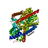
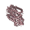
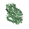
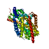
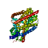
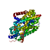
| Title | Crystal structure of LeuT bound to L-leucine in space group C2 from lipid bicelles | ||||||
 Components Components | Transporter | ||||||
 Keywords Keywords | TRANSPORT PROTEIN / Leucine transporter | ||||||
| Function / homology |  Function and homology information Function and homology information | ||||||
| Biological species |   Aquifex aeolicus (bacteria) Aquifex aeolicus (bacteria) | ||||||
| Method |  X-RAY DIFFRACTION / SYNCHROTRON / MOLECULAR REPLACEMENT / Resolution: 2.502 Å X-RAY DIFFRACTION / SYNCHROTRON / MOLECULAR REPLACEMENT / Resolution: 2.502 Å | ||||||
 Authors Authors | Wang, H. / Elferich, J. / Gouaux, E. | ||||||
 Citation Citation | Journal: Nat.Struct.Mol.Biol. / Year: 2012 Title: Structures of LeuT in bicelles define conformation and substrate binding in a membrane-like context. Authors: Wang, H. / Elferich, J. / Gouaux, E. | ||||||
| History |
|
- Structure visualization
| Structure viewer | Molecule: MolmilJmol/JSmol |
|---|
- Downloads & links
-Download
| PDBx/mmCIF format | 3usg.cif.gz | 114.3 KB | Display | PDBx/mmCIF format |
|---|---|---|---|---|
| PDB format | pdb3usg.ent.gz | 87.1 KB | Display | PDB format |
| PDBx/mmJSON format | 3usg.json.gz | Tree view | PDBx/mmJSON format | |
| Others |  Other downloads Other downloads |
-Validation report
| Arichive directory | https://data.pdbj.org/pub/pdb/validation_reports/us/3usgftp://data.pdbj.org/pub/pdb/validation_reports/us/3usg | HTTPS FTP |
|---|
-Related structure data
| Related structure data |  3usiC  3usjC  3uskC  3uslC  3usmC  3usoC  3uspC  2a65S C: citing same article ( S: Starting model for refinement |
|---|---|
| Similar structure data |

-Links
 PDBj
PDBj


- Assembly
| Deposited unit | 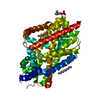
| ||||||||
|---|---|---|---|---|---|---|---|---|---|
| 1 |
| ||||||||
| 2 | 
| ||||||||
| Unit cell |
|
-Components
-Protein , 1 types, 1 molecules A
| #1: Protein | Mass: 58077.438 Da / Num. of mol.: 1 Source method: isolated from a genetically manipulated source Source: (gene. exp.) Aquifex aeolicus (bacteria) / Gene: snf, aq_2077 / Plasmid: pet16b / Production host: |
|---|
-Non-polymers , 7 types, 70 molecules 


| #2: Chemical | ChemComp-LEU /  Type: L-peptide linking / Mass: 131.173 Da / Num. of mol.: 1 / Source method: obtained synthetically / Formula: C6H13NO2 Type: L-peptide linking / Mass: 131.173 Da / Num. of mol.: 1 / Source method: obtained synthetically / Formula: C6H13NO2 | ||||||||||
|---|---|---|---|---|---|---|---|---|---|---|---|
| #3: Chemical |  Mass: 22.990 Da / Num. of mol.: 2 / Source method: obtained synthetically / Formula: Na Mass: 22.990 Da / Num. of mol.: 2 / Source method: obtained synthetically / Formula: Na#4: Chemical | ChemComp-ACT / |  Mass: 59.044 Da / Num. of mol.: 1 / Source method: obtained synthetically / Formula: C2H3O2 Mass: 59.044 Da / Num. of mol.: 1 / Source method: obtained synthetically / Formula: C2H3O2#5: Chemical | ChemComp-PEG / |  Mass: 106.120 Da / Num. of mol.: 1 / Source method: obtained synthetically / Formula: C4H10O3 Mass: 106.120 Da / Num. of mol.: 1 / Source method: obtained synthetically / Formula: C4H10O3#6: Chemical | ChemComp-UND / |  Mass: 156.308 Da / Num. of mol.: 1 / Source method: obtained synthetically / Formula: C11H24 Mass: 156.308 Da / Num. of mol.: 1 / Source method: obtained synthetically / Formula: C11H24#7: Chemical | ChemComp-PC / |  Mass: 184.151 Da / Num. of mol.: 1 / Source method: obtained synthetically / Formula: C5H15NO4P Mass: 184.151 Da / Num. of mol.: 1 / Source method: obtained synthetically / Formula: C5H15NO4P#8: Water | ChemComp-HOH / | Mass: 18.015 Da / Num. of mol.: 63 / Source method: isolated from a natural source / Formula: H2O |
-Experimental details
-Experiment
| Experiment | Method: X-RAY DIFFRACTION / Number of used crystals: 1 |
|---|
- Sample preparation
| Crystal | Density Matthews: 3.84 Å3/Da / Density % sol: 67.97 % |
|---|---|
| Crystal grow | Temperature: 293 K / Method: vapor diffusion, hanging drop / pH: 4.7 Details: 100 mM NaAc, 30-35% MPD, 5-10%PEG, pH 4.7, VAPOR DIFFUSION, HANGING DROP, temperature 293K |
-Data collection
| Diffraction | Mean temperature: 100 K |
|---|---|
| Diffraction source | Source: SYNCHROTRON / Site: APS  / Beamline: 24-ID-E / Wavelength: 0.979 Å / Beamline: 24-ID-E / Wavelength: 0.979 Å |
| Detector | Type: ADSC QUANTUM 315 / Detector: CCD / Date: Apr 25, 2011 |
| Radiation | Monochromator: Cryogenically-cooled single crystal / Protocol: SINGLE WAVELENGTH / Monochromatic (M) / Laue (L): M / Scattering type: x-ray |
| Radiation wavelength | Wavelength: 0.979 Å / Relative weight: 1 |
| Reflection | Resolution: 2.5→40 Å / Num. all: 74946 / Num. obs: 29352 / % possible obs: 97.4 % / Observed criterion σ(F): 0 / Observed criterion σ(I): 0 / Redundancy: 2.6 % / Rmerge(I) obs: 0.08 / Net I/σ(I): 10.2 |
| Reflection shell | Resolution: 2.5→2.59 Å / Redundancy: 2.4 % / Rmerge(I) obs: 0.32 / Mean I/σ(I) obs: 2 / % possible all: 90.4 |
- Processing
| Software |
| |||||||||||||||||||||||||||||||||||||||||||||||||||||||||||||||||||||||||||||
|---|---|---|---|---|---|---|---|---|---|---|---|---|---|---|---|---|---|---|---|---|---|---|---|---|---|---|---|---|---|---|---|---|---|---|---|---|---|---|---|---|---|---|---|---|---|---|---|---|---|---|---|---|---|---|---|---|---|---|---|---|---|---|---|---|---|---|---|---|---|---|---|---|---|---|---|---|---|---|
| Refinement | Method to determine structure: MOLECULAR REPLACEMENT Starting model: pdb entry 2A65 Resolution: 2.502→36.674 Å / SU ML: 0.3 / σ(F): 0 / Phase error: 25.18 / Stereochemistry target values: ML
| |||||||||||||||||||||||||||||||||||||||||||||||||||||||||||||||||||||||||||||
| Solvent computation | Shrinkage radii: 0.49 Å / VDW probe radii: 0.8 Å / Solvent model: FLAT BULK SOLVENT MODEL / Bsol: 53.26 Å2 / ksol: 0.32 e/Å3 | |||||||||||||||||||||||||||||||||||||||||||||||||||||||||||||||||||||||||||||
| Displacement parameters |
| |||||||||||||||||||||||||||||||||||||||||||||||||||||||||||||||||||||||||||||
| Refinement step | Cycle: LAST / Resolution: 2.502→36.674 Å
| |||||||||||||||||||||||||||||||||||||||||||||||||||||||||||||||||||||||||||||
| Refine LS restraints |
| |||||||||||||||||||||||||||||||||||||||||||||||||||||||||||||||||||||||||||||
| LS refinement shell |
|
