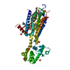
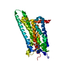
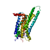
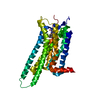
| Entry | Database: PDB / ID: 3pwh
|
|---|
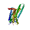
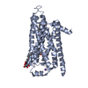
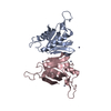
| Title | Thermostabilised Adenosine A2A Receptor |
|---|
 Components Components | Adenosine receptor A2a |
|---|
 Keywords Keywords | SIGNALING PROTEIN / 7TM / GPCR / inverse agonist / G-protein / Membrane Protein |
|---|
| Function / homology |  Function and homology information Function and homology information
regulation of norepinephrine secretion / positive regulation of acetylcholine secretion, neurotransmission / negative regulation of alpha-beta T cell activation / positive regulation of circadian sleep/wake cycle, sleep / Adenosine P1 receptors / G protein-coupled adenosine receptor activity / response to purine-containing compound / G protein-coupled adenosine receptor signaling pathway / NGF-independant TRKA activation / Surfactant metabolism ...regulation of norepinephrine secretion / positive regulation of acetylcholine secretion, neurotransmission / negative regulation of alpha-beta T cell activation / positive regulation of circadian sleep/wake cycle, sleep / Adenosine P1 receptors / G protein-coupled adenosine receptor activity / response to purine-containing compound / G protein-coupled adenosine receptor signaling pathway / NGF-independant TRKA activation / Surfactant metabolism / sensory perception / synaptic transmission, dopaminergic / type 5 metabotropic glutamate receptor binding / negative regulation of vascular permeability / synaptic transmission, cholinergic / intermediate filament / positive regulation of glutamate secretion / presynaptic active zone / positive regulation of urine volume / blood circulation / response to caffeine / eating behavior / inhibitory postsynaptic potential / alpha-actinin binding / regulation of calcium ion transport / asymmetric synapse / axolemma / membrane depolarization / prepulse inhibition / cellular defense response / phagocytosis / positive regulation of synaptic transmission, glutamatergic / neuron projection morphogenesis / astrocyte activation / presynaptic modulation of chemical synaptic transmission / central nervous system development / response to amphetamine / positive regulation of long-term synaptic potentiation / positive regulation of synaptic transmission, GABAergic / positive regulation of protein secretion / positive regulation of apoptotic signaling pathway / regulation of mitochondrial membrane potential / excitatory postsynaptic potential / synaptic transmission, glutamatergic / apoptotic signaling pathway / locomotory behavior / negative regulation of inflammatory response / vasodilation / adenylate cyclase-modulating G protein-coupled receptor signaling pathway / adenylate cyclase-activating G protein-coupled receptor signaling pathway / blood coagulation / cell-cell signaling / presynaptic membrane / G alpha (s) signalling events / phospholipase C-activating G protein-coupled receptor signaling pathway / negative regulation of neuron apoptotic process / postsynaptic membrane / calmodulin binding / positive regulation of ERK1 and ERK2 cascade / response to xenobiotic stimulus / inflammatory response / negative regulation of cell population proliferation / neuronal cell body / apoptotic process / dendrite / regulation of DNA-templated transcription / lipid binding / protein-containing complex binding / glutamatergic synapse / enzyme binding / identical protein binding / membrane / plasma membraneSimilarity search - Function Adenosine A2A receptor / Adenosine receptor / Rhopdopsin 7-helix transmembrane proteins / Rhodopsin 7-helix transmembrane proteins / Serpentine type 7TM GPCR chemoreceptor Srsx / G-protein coupled receptors family 1 signature. / 7 transmembrane receptor (rhodopsin family) / G protein-coupled receptor, rhodopsin-like / GPCR, rhodopsin-like, 7TM / G-protein coupled receptors family 1 profile. ...Adenosine A2A receptor / Adenosine receptor / Rhopdopsin 7-helix transmembrane proteins / Rhodopsin 7-helix transmembrane proteins / Serpentine type 7TM GPCR chemoreceptor Srsx / G-protein coupled receptors family 1 signature. / 7 transmembrane receptor (rhodopsin family) / G protein-coupled receptor, rhodopsin-like / GPCR, rhodopsin-like, 7TM / G-protein coupled receptors family 1 profile. / Up-down Bundle / Mainly AlphaSimilarity search - Domain/homology |
|---|
| Biological species |  Homo sapiens (human) Homo sapiens (human) |
|---|
| Method |  X-RAY DIFFRACTION / SYNCHROTRON / MOLECULAR REPLACEMENT / Resolution: 3.296 Å X-RAY DIFFRACTION / SYNCHROTRON / MOLECULAR REPLACEMENT / Resolution: 3.296 Å |
|---|
 Authors Authors | Dore, A.S. / Robertson, N. / Errey, J.C. / Ng, I. / Tehan, B. / Hurrell, E. / Magnani, F. / Tate, C.G. / Weir, M. / Marshall, F.H. |
|---|
 Citation Citation | Journal: Structure / Year: 2011
Title: Structure of the adenosine A(2A) receptor in complex with ZM241385 and the xanthines XAC and caffeine
Authors: Dore, A.S. / Robertson, N. / Errey, J.C. / Ng, I. / Hollenstein, K. / Tehan, B. / Hurrell, E. / Bennett, K. / Congreve, M. / Magnani, F. / Tate, C.G. / Weir, M. / Marshall, F.H. |
|---|
| History | | Deposition | Dec 8, 2010 | Deposition site: RCSB / Processing site: PDBJ |
|---|
| Revision 1.0 | Sep 7, 2011 | Provider: repository / Type: Initial release |
|---|
| Revision 1.1 | Jun 20, 2012 | Group: Database references |
|---|
| Revision 1.2 | Nov 1, 2023 | Group: Data collection / Database references ...Data collection / Database references / Derived calculations / Refinement description
Category: chem_comp_atom / chem_comp_bond ...chem_comp_atom / chem_comp_bond / database_2 / pdbx_initial_refinement_model / struct_ref_seq_dif / struct_site
Item: _database_2.pdbx_DOI / _database_2.pdbx_database_accession ..._database_2.pdbx_DOI / _database_2.pdbx_database_accession / _struct_ref_seq_dif.details / _struct_site.pdbx_auth_asym_id / _struct_site.pdbx_auth_comp_id / _struct_site.pdbx_auth_seq_id |
|---|
| Revision 1.3 | Nov 20, 2024 | Group: Structure summary / Category: pdbx_entry_details / pdbx_modification_feature |
|---|
|
|---|
 Movie
Movie Controller


 Open data
 Basic information
 Structure visualization
 Downloads & links
 Other downloads


 PDBj
PDBj


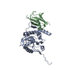
 Assembly
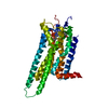

 Spodoptera frugiperda (fall armyworm) / References: UniProt: P29274
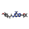
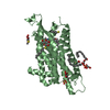
 Mass: 337.336 Da / Num. of mol.: 1 / Source method: obtained synthetically / Formula: C16H15N7O2 / Comment: antagonist*YM
 Sample preparation
 / Beamline: I24 / Wavelength: 0.9777 Å
 Processing