 Movie
Movie Controller

[English] 日本語
 Yorodumi
- PDB-3pcn: STRUCTURE OF PROTOCATECHUATE 3,4-DIOXYGENASE COMPLEXED WITH 3,4-D... -
+ Open data

- Basic information
| Entry | Database: PDB / ID: 3pcn | |||||||||
|---|---|---|---|---|---|---|---|---|---|---|
| Title | STRUCTURE OF PROTOCATECHUATE 3,4-DIOXYGENASE COMPLEXED WITH 3,4-DIHYDROXYPHENYLACETATE | |||||||||
 Components Components | (PROTOCATECHUATE 3,4- ...) x 2 | |||||||||
 Keywords Keywords | DIOXYGENASE / IRON / NONHEME / METALLOPROTEIN / OXIDOREDUCTASE / SUBSTRATE COMPLEX | |||||||||
| Function / homology |  Function and homology information Function and homology informationprotocatechuate 3,4-dioxygenase / protocatechuate 3,4-dioxygenase activity / 3,4-dihydroxybenzoate catabolic process / beta-ketoadipate pathway / ferric iron binding Similarity search - Function | |||||||||
| Biological species |  Pseudomonas putida (bacteria) Pseudomonas putida (bacteria) | |||||||||
| Method |  X-RAY DIFFRACTION / NATIVE MODEL PHASES / Resolution: 2.4 Å X-RAY DIFFRACTION / NATIVE MODEL PHASES / Resolution: 2.4 Å | |||||||||
 Authors Authors | Orville, A.M. / Lipscomb, J.D. / Ohlendorf, D.H. | |||||||||
 Citation Citation | Journal: Biochemistry / Year: 1997 Title: Crystal structure and resonance Raman studies of protocatechuate 3,4-dioxygenase complexed with 3,4-dihydroxyphenylacetate. Authors: Elgren, T.E. / Orville, A.M. / Kelly, K.A. / Lipscomb, J.D. / Ohlendorf, D.H. / Que Jr., L. #1: Journal: Biochemistry / Year: 1997Title: Crystal Structures of Substrate and Substrate Analog Complexes of Protocatechuate 3,4-Dioxygenase: Endogenous Fe3+ Ligand Displacement in Response to Substrate Binding Authors: Orville, A.M. / Lipscomb, J.D. / Ohlendorf, D.H. #2: Journal: Biochemistry / Year: 1997Title: Structures of Competitive Inhibitor Complexes of Protocatechuate 3,4-Dioxygenase: Multiple Exogenous Ligand Binding Orientations within the Active Site Authors: Orville, A.M. / Elango, N. / Lipscomb, J.D. / Ohlendorf, D.H. #3: Journal: J.Mol.Biol. / Year: 1994Title: Structure of Protocatechuate 3,4-Dioxygenase from Pseudomonas Aeruginosa at 2.15 A Resolution Authors: Ohlendorf, D.H. / Orville, A.M. / Lipscomb, J.D. #4: Journal: Nature / Year: 1988Title: Structure and Assembly of Protocatechuate 3,4-Dioxygenase Authors: Ohlendorf, D.H. / Lipscomb, J.D. / Weber, P.C. | |||||||||
| History |
|
- Structure visualization
| Structure viewer | Molecule: MolmilJmol/JSmol |
|---|
- Downloads & links
-Download
| PDBx/mmCIF format | 3pcn.cif.gz | 539 KB | Display | PDBx/mmCIF format |
|---|---|---|---|---|
| PDB format | pdb3pcn.ent.gz | 439.8 KB | Display | PDB format |
| PDBx/mmJSON format | 3pcn.json.gz | Tree view | PDBx/mmJSON format | |
| Others |  Other downloads Other downloads |
-Validation report
| Arichive directory | https://data.pdbj.org/pub/pdb/validation_reports/pc/3pcnftp://data.pdbj.org/pub/pdb/validation_reports/pc/3pcn | HTTPS FTP |
|---|
-Related structure data
| Similar structure data |
|---|

-Links
 PDBj
PDBj


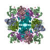
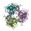
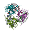
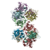
- Assembly
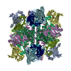
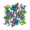
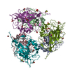
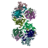
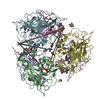
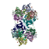
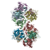
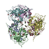
| Deposited unit | 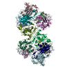
| ||||||||||||||||||
|---|---|---|---|---|---|---|---|---|---|---|---|---|---|---|---|---|---|---|---|
| 1 | 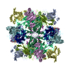
| ||||||||||||||||||
| Unit cell |
| ||||||||||||||||||
| Noncrystallographic symmetry (NCS) | NCS oper:
| ||||||||||||||||||
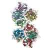
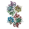
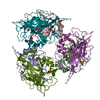
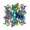
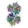
| Details | THE ACTIVE FORM OF THE ENZYME IS THE WHOLE UNIT CELL CONTENT. IT CONSISTS OF 12 PROTOMERS. EACH PROTOMER IS MADE OF TWO POLYPEPTIDE CHAINS AND ONE FE(III). THE ASYMMETRIC UNIT CONSISTS OF 6 PROTOMERS. THEY ARE: CHAINS (A, M), CHAINS (B, N), CHAINS (C, O), CHAINS (D, P), CHAINS (E, Q), CHAINS (F, R). EACH PROTOMER CONTAINS AN ACTIVE SITE WITH FE(III) AS THE CENTER AND A VESTIGIAL SITE. THE ACTIVE SITES ARE NAMED: ACA, ACB, ACC, ACD, ACE, ACF. THE VESTIGIAL SITES ARE NAMED VEA, VEB, VEC, VED, VEE AND VEF. THE CHAIN IDENTIFIERS HAVE BEEN ASSIGNED AS FOLLOWS: ALPHA BETA WATERS A M G B N H C O I D P J E Q K F R L |
-Components
-PROTOCATECHUATE 3,4- ... , 2 types, 12 molecules ABCDEFMNOPQR
| #1: Protein | Mass: 22278.812 Da / Num. of mol.: 6 / Source method: isolated from a natural source / Details: PREVIOUSLY CLASSIFIED AS PSEUDOMONAS AERUGINOSA / Source: (natural) Pseudomonas putida (bacteria)References: UniProt: P00436, protocatechuate 3,4-dioxygenase #2: Protein | Mass: 26696.287 Da / Num. of mol.: 6 / Source method: isolated from a natural source / Details: PREVIOUSLY CLASSIFIED AS PSEUDOMONAS AERUGINOSA / Source: (natural) Pseudomonas putida (bacteria)References: UniProt: P00437, protocatechuate 3,4-dioxygenase |
|---|
-Non-polymers , 4 types, 1392 molecules 

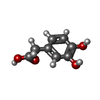

| #3: Chemical | ChemComp-FE /  Mass: 55.845 Da / Num. of mol.: 6 / Source method: obtained synthetically / Formula: Fe Mass: 55.845 Da / Num. of mol.: 6 / Source method: obtained synthetically / Formula: Fe#4: Chemical | ChemComp-BME /  Mass: 78.133 Da / Num. of mol.: 6 / Source method: obtained synthetically / Formula: C2H6OS Mass: 78.133 Da / Num. of mol.: 6 / Source method: obtained synthetically / Formula: C2H6OS#5: Chemical | ChemComp-DHY /  Mass: 168.147 Da / Num. of mol.: 6 / Source method: obtained synthetically / Formula: C8H8O4 / Comment: neurotransmitter*YM Mass: 168.147 Da / Num. of mol.: 6 / Source method: obtained synthetically / Formula: C8H8O4 / Comment: neurotransmitter*YM#6: Water | ChemComp-HOH / | Mass: 18.015 Da / Num. of mol.: 1374 / Source method: isolated from a natural source / Formula: H2O |
|---|
-Details
| Nonpolymer details | THE SUBSTRATE, DHY 550, FITS THE ELECTRON DENSITY EQUALLY WELL IN EITHER OF TWO ORIENTATIONS AND IS ...THE SUBSTRATE, DHY 550, FITS THE ELECTRON DENSITY EQUALLY WELL IN EITHER OF TWO ORIENTATIO |
|---|
-Experimental details
-Experiment
| Experiment | Method: X-RAY DIFFRACTION / Number of used crystals: 2 |
|---|
- Sample preparation
| Crystal | Density Matthews: 2.84 Å3/Da / Density % sol: 56.63 % | |||||||||||||||||||||||||
|---|---|---|---|---|---|---|---|---|---|---|---|---|---|---|---|---|---|---|---|---|---|---|---|---|---|---|
| Crystal grow | pH: 8.4 / Details: pH 8.4 | |||||||||||||||||||||||||
| Crystal grow | *PLUS Temperature: 4 ℃ / Method: vapor diffusion, hanging drop / Details: Ohlendorf, D.H., (1994) J.Mol.Biol., 244, 586. | |||||||||||||||||||||||||
| Components of the solutions | *PLUS
|
-Data collection
| Diffraction | Mean temperature: 296 K |
|---|---|
| Diffraction source | Source: ROTATING ANODE / Type: RIGAKU RUH2R / Wavelength: 1.5418 |
| Detector | Type: SIEMENS / Detector: AREA DETECTOR / Date: Oct 11, 1994 / Details: COLLIMATOR |
| Radiation | Monochromator: GRAPHITE(002) / Monochromatic (M) / Laue (L): M / Scattering type: x-ray |
| Radiation wavelength | Wavelength: 1.5418 Å / Relative weight: 1 |
| Reflection | Resolution: 2.4→10 Å / Num. obs: 127824 / % possible obs: 97 % / Observed criterion σ(I): 1 / Redundancy: 6.2 % / Rsym value: 0.11 / Net I/σ(I): 9.75 |
| Reflection shell | Resolution: 2.4→2.5 Å / Redundancy: 3.5 % / Mean I/σ(I) obs: 1.3 / % possible all: 89 |
| Reflection | *PLUS Num. measured all: 792401 / Rmerge(I) obs: 0.11 |
- Processing
| Software |
| ||||||||||||||||||||||||||||||||||||||||||||||||||||||||||||||||||||||||||||||||||||
|---|---|---|---|---|---|---|---|---|---|---|---|---|---|---|---|---|---|---|---|---|---|---|---|---|---|---|---|---|---|---|---|---|---|---|---|---|---|---|---|---|---|---|---|---|---|---|---|---|---|---|---|---|---|---|---|---|---|---|---|---|---|---|---|---|---|---|---|---|---|---|---|---|---|---|---|---|---|---|---|---|---|---|---|---|---|
| Refinement | Method to determine structure: NATIVE MODEL PHASES / Resolution: 2.4→6 Å / σ(F): 1 Details: THE NONSTANDARD UNIT CELL I 2 WAS CHOSEN OVER THE EQUIVALENT C 2 CELL BECAUSE OF THE CONVENIENCE OF HAVING A BETA ANGLE NEAR 90 DEGREES AND TO MAINTAIN CONSISTENCY WITH THE INITIAL ...Details: THE NONSTANDARD UNIT CELL I 2 WAS CHOSEN OVER THE EQUIVALENT C 2 CELL BECAUSE OF THE CONVENIENCE OF HAVING A BETA ANGLE NEAR 90 DEGREES AND TO MAINTAIN CONSISTENCY WITH THE INITIAL CRYSTALLIZATION REPORT. TO CONVERT FROM FRACTIONAL I 2 TO FRACTIONAL C 2, USE THE FOLLOWING TRANSFORMATION: [ 1.0 ][ 0.0 ][ 0.0 ] (XFRAC_I2) (XFRAC_C2) [ 0.0 ][ 1.0 ][ 0.0 ] X (YFRAC_I2) = (YFRAC_C2) [ -1.0 ][ 0.0 ][ 1.0 ] (ZFRAC_I2) (ZFRAC_C2) THE ORTHOGONAL COORDINATE SYSTEM CHOSEN FOR THIS ENTRY IS THAT DEFINED BY THE LOCAL SYMMETRY OF THE COMPLEX (23).
| ||||||||||||||||||||||||||||||||||||||||||||||||||||||||||||||||||||||||||||||||||||
| Displacement parameters | Biso mean: 21.93 Å2 | ||||||||||||||||||||||||||||||||||||||||||||||||||||||||||||||||||||||||||||||||||||
| Refinement step | Cycle: LAST / Resolution: 2.4→6 Å
| ||||||||||||||||||||||||||||||||||||||||||||||||||||||||||||||||||||||||||||||||||||
| Refine LS restraints |
| ||||||||||||||||||||||||||||||||||||||||||||||||||||||||||||||||||||||||||||||||||||
| Software | *PLUS Name: PROLSQ / Classification: refinement | ||||||||||||||||||||||||||||||||||||||||||||||||||||||||||||||||||||||||||||||||||||
| Refinement | *PLUS Rfactor obs: 0.166 | ||||||||||||||||||||||||||||||||||||||||||||||||||||||||||||||||||||||||||||||||||||
| Solvent computation | *PLUS | ||||||||||||||||||||||||||||||||||||||||||||||||||||||||||||||||||||||||||||||||||||
| Displacement parameters | *PLUS Biso mean: 21.938 Å2 | ||||||||||||||||||||||||||||||||||||||||||||||||||||||||||||||||||||||||||||||||||||
| LS refinement shell | *PLUS Highest resolution: 2.4 Å / Lowest resolution: 2.6 Å / Rfactor obs: 0.209 |
