 Movie
Movie Controller

[English] 日本語
 Yorodumi
- PDB-3mi5: Axial Ligand Swapping In Double Mutant Maintains Intradiol-cleava... -
+ Open data

- Basic information
| Entry | Database: PDB / ID: 3mi5 | ||||||
|---|---|---|---|---|---|---|---|
| Title | Axial Ligand Swapping In Double Mutant Maintains Intradiol-cleavage Chemistry in Protocatechuate 3,4-Dioxygenase | ||||||
 Components Components | (Protocatechuate 3,4-dioxygenase ...) x 2 | ||||||
 Keywords Keywords | OXIDOREDUCTASE / dioxygenase / non-heme / iron / intradiol / catechol / substrate analogue / protocatechuate | ||||||
| Function / homology |  Function and homology information Function and homology informationprotocatechuate 3,4-dioxygenase / protocatechuate 3,4-dioxygenase activity / 3,4-dihydroxybenzoate catabolic process / beta-ketoadipate pathway / ferric iron binding Similarity search - Function | ||||||
| Biological species |  Pseudomonas putida (bacteria) Pseudomonas putida (bacteria) | ||||||
| Method |  X-RAY DIFFRACTION / SYNCHROTRON / MOLECULAR REPLACEMENT / molecular replacement / Resolution: 1.78 Å X-RAY DIFFRACTION / SYNCHROTRON / MOLECULAR REPLACEMENT / molecular replacement / Resolution: 1.78 Å | ||||||
 Authors Authors | Purpero, V.M. / Lipscomb, J.D. | ||||||
 Citation Citation | Journal: To be Published Title: Axial Ligand Swapping In Double Mutant Maintains Intradiol-cleavage Chemistry in Protocatechuate 3,4-Dioxygenase Authors: Purpero, V.M. / Lipscomb, J.D. | ||||||
| History |
|
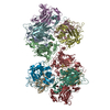
- Structure visualization
| Structure viewer | Molecule: MolmilJmol/JSmol |
|---|
- Downloads & links
-Download
| PDBx/mmCIF format | 3mi5.cif.gz | 595.4 KB | Display | PDBx/mmCIF format |
|---|---|---|---|---|
| PDB format | pdb3mi5.ent.gz | 487.1 KB | Display | PDB format |
| PDBx/mmJSON format | 3mi5.json.gz | Tree view | PDBx/mmJSON format | |
| Others |  Other downloads Other downloads |
-Validation report
| Arichive directory | https://data.pdbj.org/pub/pdb/validation_reports/mi/3mi5ftp://data.pdbj.org/pub/pdb/validation_reports/mi/3mi5 | HTTPS FTP |
|---|
-Related structure data
| Related structure data | 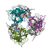 3mflC  3mi1C  3mv4C 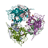 3mv6C  3t63C  3t67C  3ini C: citing same article ( |
|---|---|
| Similar structure data |

-Links
 PDBj
PDBj


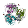
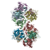
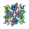
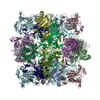
- Assembly
| Deposited unit | 
| ||||||||
|---|---|---|---|---|---|---|---|---|---|
| 1 | 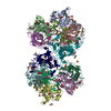
| ||||||||
| Unit cell |
| ||||||||
| Components on special symmetry positions |
| ||||||||
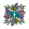
| Details | exists as a dodecamer (12) of dimer in solution. The space group C2 shows 6 of 12 (x,y,z). By applying the symmetry operator (-x,-y,-z) this completes the biological unit assembly. |
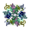
-Components
-Protocatechuate 3,4-dioxygenase ... , 2 types, 12 molecules ABCDEFMNOPQR
| #1: Protein | Mass: 22278.812 Da / Num. of mol.: 6 Source method: isolated from a genetically manipulated source Source: (gene. exp.) Pseudomonas putida (bacteria) / Strain: P. putida / Gene: pcaG / Plasmid: pCE vector, pT7-7 / Production host: References: UniProt: P00436, protocatechuate 3,4-dioxygenase #2: Protein | Mass: 26696.287 Da / Num. of mol.: 6 / Mutation: Y148H/H163Y Source method: isolated from a genetically manipulated source Source: (gene. exp.) Pseudomonas putida (bacteria) / Gene: pcaH / Plasmid: pCE vector, pT7-7 / Production host: References: UniProt: P00437, protocatechuate 3,4-dioxygenase |
|---|
-Non-polymers , 7 types, 2402 molecules 


| #3: Chemical | ChemComp-SO4 /  Mass: 96.063 Da / Num. of mol.: 12 / Source method: obtained synthetically / Formula: SO4 Mass: 96.063 Da / Num. of mol.: 12 / Source method: obtained synthetically / Formula: SO4#4: Chemical | ChemComp-BME /  Mass: 78.133 Da / Num. of mol.: 25 / Source method: obtained synthetically / Formula: C2H6OS Mass: 78.133 Da / Num. of mol.: 25 / Source method: obtained synthetically / Formula: C2H6OS#5: Chemical | ChemComp-GOL /  Mass: 92.094 Da / Num. of mol.: 42 / Source method: obtained synthetically / Formula: C3H8O3 Mass: 92.094 Da / Num. of mol.: 42 / Source method: obtained synthetically / Formula: C3H8O3#6: Chemical | ChemComp-CAQ /  Mass: 110.111 Da / Num. of mol.: 12 / Source method: obtained synthetically / Formula: C6H6O2 Mass: 110.111 Da / Num. of mol.: 12 / Source method: obtained synthetically / Formula: C6H6O2#7: Chemical | ChemComp-FE /  Mass: 55.845 Da / Num. of mol.: 6 / Source method: obtained synthetically / Formula: Fe Mass: 55.845 Da / Num. of mol.: 6 / Source method: obtained synthetically / Formula: Fe#8: Chemical | ChemComp-CL /  Mass: 35.453 Da / Num. of mol.: 5 / Source method: obtained synthetically / Formula: Cl Mass: 35.453 Da / Num. of mol.: 5 / Source method: obtained synthetically / Formula: Cl#9: Water | ChemComp-HOH / | Mass: 18.015 Da / Num. of mol.: 2300 / Source method: isolated from a natural source / Formula: H2O |
|---|
-Experimental details
-Experiment
| Experiment | Method: X-RAY DIFFRACTION / Number of used crystals: 1 |
|---|
- Sample preparation
| Crystal | Density Matthews: 2.58 Å3/Da / Density % sol: 52.34 % |
|---|---|
| Crystal grow | Temperature: 277 K / Method: vapor diffusion, hanging drop / pH: 8.5 Details: 1.5-1.8 M ammounium sulfate, 40-60 mM TRIS pH 8.5, 5 mM BME protein conc. 15-25 mg/mL, VAPOR DIFFUSION, HANGING DROP, temperature 277K |
-Data collection
| Diffraction | Mean temperature: 100 K |
|---|---|
| Diffraction source | Source: SYNCHROTRON / Site: APS  / Beamline: 19-BM / Wavelength: 0.98 Å / Beamline: 19-BM / Wavelength: 0.98 Å |
| Detector | Type: ADSC QUANTUM 210r / Detector: CCD / Date: Apr 15, 2009 / Details: mirrors |
| Radiation | Monochromator: double crystal / Protocol: SINGLE WAVELENGTH / Monochromatic (M) / Laue (L): M / Scattering type: x-ray |
| Radiation wavelength | Wavelength: 0.98 Å / Relative weight: 1 |
| Reflection | Resolution: 1.78→50 Å / Num. all: 284851 / Num. obs: 282736 / % possible obs: 99.3 % / Observed criterion σ(F): 1 / Observed criterion σ(I): 5 / Redundancy: 3 % / Biso Wilson estimate: 19.77 Å2 / Rmerge(I) obs: 0.058 / Χ2: 1.16 / Net I/σ(I): 15.5 |
| Reflection shell | Resolution: 1.78→1.81 Å / Redundancy: 2.4 % / Rmerge(I) obs: 0.37 / Mean I/σ(I) obs: 2.76 / Num. unique all: 14065 / Χ2: 1.111 / % possible all: 99.2 |
-Phasing
| Phasing | Method: molecular replacement |
|---|
- Processing
| Software |
| |||||||||||||||||||||||||||||||||||||||||||||||||||||||||||||||||
|---|---|---|---|---|---|---|---|---|---|---|---|---|---|---|---|---|---|---|---|---|---|---|---|---|---|---|---|---|---|---|---|---|---|---|---|---|---|---|---|---|---|---|---|---|---|---|---|---|---|---|---|---|---|---|---|---|---|---|---|---|---|---|---|---|---|---|
| Refinement | Method to determine structure: MOLECULAR REPLACEMENT / Resolution: 1.78→30.62 Å / Cor.coef. Fo:Fc: 0.97 / Cor.coef. Fo:Fc free: 0.955 / WRfactor Rfree: 0.182 / WRfactor Rwork: 0.15 / Occupancy max: 1 / Occupancy min: 0 / FOM work R set: 0.89 / SU B: 2.009 / SU ML: 0.064 / SU R Cruickshank DPI: 0.103 / SU Rfree: 0.101 / Cross valid method: THROUGHOUT / σ(F): 1 / σ(I): 3 / ESU R: 0.103 / ESU R Free: 0.101 / Stereochemistry target values: MAXIMUM LIKELIHOOD Details: HYDROGENS HAVE BEEN ADDED IN THE RIDING POSITIONS U VALUES : REFINED INDIVIDUALLY
| |||||||||||||||||||||||||||||||||||||||||||||||||||||||||||||||||
| Solvent computation | Ion probe radii: 0.8 Å / Shrinkage radii: 0.8 Å / VDW probe radii: 1.4 Å / Solvent model: BABINET MODEL WITH MASK | |||||||||||||||||||||||||||||||||||||||||||||||||||||||||||||||||
| Displacement parameters | Biso max: 70.21 Å2 / Biso mean: 21.401 Å2 / Biso min: 4.69 Å2
| |||||||||||||||||||||||||||||||||||||||||||||||||||||||||||||||||
| Refinement step | Cycle: LAST / Resolution: 1.78→30.62 Å
| |||||||||||||||||||||||||||||||||||||||||||||||||||||||||||||||||
| Refine LS restraints |
| |||||||||||||||||||||||||||||||||||||||||||||||||||||||||||||||||
| LS refinement shell | Resolution: 1.777→1.823 Å / Total num. of bins used: 20
|
