 Movie
Movie Controller

[English] 日本語
 Yorodumi
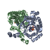
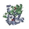
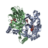
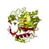
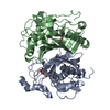
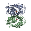
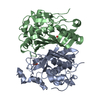
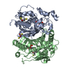
- PDB-3mf2: Crystal structure of class II aaRS homologue (Bll0957) complexed ... -
+ Open data

- Basic information
| Entry | Database: PDB / ID: 3mf2 | ||||||
|---|---|---|---|---|---|---|---|
| Title | Crystal structure of class II aaRS homologue (Bll0957) complexed with AMP | ||||||
 Components Components | Bll0957 protein | ||||||
 Keywords Keywords |  LIGASE / aminoacyl-tRNA synthetase / seryl-tRNA synthetase / zinc ion / amino acid:[carrier protein] ligase / Bll0957 LIGASE / aminoacyl-tRNA synthetase / seryl-tRNA synthetase / zinc ion / amino acid:[carrier protein] ligase / Bll0957 | ||||||
| Function / homology |  Function and homology information Function and homology information: / aminoacyl-tRNA ligase activity / tRNA aminoacylation for protein translation / ATP binding / metal ion bindingSimilarity search - Function | ||||||
| Biological species |  Bradyrhizobium japonicum (bacteria) Bradyrhizobium japonicum (bacteria) | ||||||
| Method | X-RAY DIFFRACTION / SYNCHROTRON / SAD / Resolution: 2.15 Å | ||||||
 Authors Authors | Weygand-Durasevic, I. / Mocibob, M. / Ivic, N. / Bilokapic, S. / Maier, T. / Luic, M. / Ban, N. | ||||||
 Citation Citation | Journal: Proc.Natl.Acad.Sci.USA / Year: 2010 Title: Homologs of aminoacyl-tRNA synthetases acylate carrier proteins and provide a link between ribosomal and nonribosomal peptide synthesis Authors: Mocibob, M. / Ivic, N. / Bilokapic, S. / Maier, T. / Luic, M. / Ban, N. / Weygand-Durasevic, I. #1: Journal: Embo J. / Year: 2006Title: Structure of the unusual seryl-tRNA synthetase reveals a distinct zinc-dependent mode of substrate recognition Authors: Bilokapic, S. / Maier, T. / Ahel, D. / Gruic-Sovulj, I. / Soll, D. / Weygand-Durasevic, I. / Ban, N. | ||||||
| History |
|
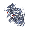
- Structure visualization
| Structure viewer | Molecule: MolmilJmol/JSmol |
|---|
- Downloads & links
-Download
| PDBx/mmCIF format | 3mf2.cif.gz | 131.3 KB | Display | PDBx/mmCIF format |
|---|---|---|---|---|
| PDB format | pdb3mf2.ent.gz | 106 KB | Display | PDB format |
| PDBx/mmJSON format | 3mf2.json.gz | Tree view | PDBx/mmJSON format | |
| Others |  Other downloads Other downloads |
-Validation report
| Arichive directory | https://data.pdbj.org/pub/pdb/validation_reports/mf/3mf2ftp://data.pdbj.org/pub/pdb/validation_reports/mf/3mf2 | HTTPS FTP |
|---|
-Related structure data


-Links
 PDBj
PDBj


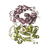
- Assembly
| Deposited unit | 
| ||||||||
|---|---|---|---|---|---|---|---|---|---|
| 1 |
| ||||||||
| Unit cell |
|
-Components
| #1: Protein | Mass: 38159.203 Da / Num. of mol.: 2 Source method: isolated from a genetically manipulated source Source: (gene. exp.) Bradyrhizobium japonicum (bacteria) / Strain: USDA110 / Gene: bll0957 / Plasmid: pET28b / Production host: Escherichia coli (E. coli) / Strain (production host): BL21(DE3) / References: UniProt: Q89VT8#2: Chemical |   Mass: 65.409 Da / Num. of mol.: 2 / Source method: obtained synthetically / Formula: Zn Mass: 65.409 Da / Num. of mol.: 2 / Source method: obtained synthetically / Formula: Zn#3: Chemical | Adenosine monophosphate  Mass: 347.221 Da / Num. of mol.: 2 / Source method: obtained synthetically / Formula: C10H14N5O7P / Comment: AMP*YM Mass: 347.221 Da / Num. of mol.: 2 / Source method: obtained synthetically / Formula: C10H14N5O7P / Comment: AMP*YM#4: Chemical | Glycerol  Mass: 92.094 Da / Num. of mol.: 2 / Source method: obtained synthetically / Formula: C3H8O3 Mass: 92.094 Da / Num. of mol.: 2 / Source method: obtained synthetically / Formula: C3H8O3#5: Water | ChemComp-HOH / | Water Mass: 18.015 Da / Num. of mol.: 170 / Source method: isolated from a natural source / Formula: H2O Mass: 18.015 Da / Num. of mol.: 170 / Source method: isolated from a natural source / Formula: H2O |
|---|
-Experimental details
-Experiment
| Experiment | Method: X-RAY DIFFRACTION / Number of used crystals: 1 |
|---|
- Sample preparation
| Crystal | Density Matthews: 2.11 Å3/Da / Density % sol: 41.59 % |
|---|---|
| Crystal grow | Temperature: 291 K / Method: hanging drop / pH: 4.6 Details: PEG 4000, gycerol, pH 4.6, hanging drop, temperature 291K |
-Data collection
| Diffraction | Mean temperature: 100 K | ||||||||||||||||||||||||||||||||||||||||||||||||||||||||||||
|---|---|---|---|---|---|---|---|---|---|---|---|---|---|---|---|---|---|---|---|---|---|---|---|---|---|---|---|---|---|---|---|---|---|---|---|---|---|---|---|---|---|---|---|---|---|---|---|---|---|---|---|---|---|---|---|---|---|---|---|---|---|
| Diffraction source | Source: SYNCHROTRON / Site: SOLEIL  / Beamline: PROXIMA 1 / Wavelength: 1.282 Å / Beamline: PROXIMA 1 / Wavelength: 1.282 Å | ||||||||||||||||||||||||||||||||||||||||||||||||||||||||||||
| Detector | Type: ADSC QUANTUM 315r / Detector: CCD / Date: Sep 20, 2008 | ||||||||||||||||||||||||||||||||||||||||||||||||||||||||||||
| Radiation | Monochromator: Si 311 / Protocol: SINGLE WAVELENGTH / Monochromatic (M) / Laue (L): M / Scattering type: x-ray | ||||||||||||||||||||||||||||||||||||||||||||||||||||||||||||
| Radiation wavelength | Wavelength: 1.282 Å / Relative weight: 1 | ||||||||||||||||||||||||||||||||||||||||||||||||||||||||||||
| Reflection | Number: 278833 / Rmerge(I) obs: 0.055 / D res high: 2.15 Å / D res low: 46.74 Å / Num. obs: 35847 / % possible obs: 99.4 | ||||||||||||||||||||||||||||||||||||||||||||||||||||||||||||
| Diffraction reflection shell |
| ||||||||||||||||||||||||||||||||||||||||||||||||||||||||||||
| Reflection | Resolution: 2.15→46.74 Å / Num. all: 35915 / Num. obs: 35847 / % possible obs: 99.4 % / Observed criterion σ(I): -3 / Biso Wilson estimate: 41.182 Å2 / Rmerge(I) obs: 0.055 / Net I/σ(I): 16.23 | ||||||||||||||||||||||||||||||||||||||||||||||||||||||||||||
| Reflection shell | Resolution: 2.15→2.28 Å / Rmerge(I) obs: 0.407 / Mean I/σ(I) obs: 3.4 / Num. measured obs: 40479 / Num. unique obs: 10638 / % possible all: 96.8 |
-Phasing
| Phasing | Method: SAD |
|---|
- Processing
| Software |
| ||||||||||||||||||||||||||||||||||||
|---|---|---|---|---|---|---|---|---|---|---|---|---|---|---|---|---|---|---|---|---|---|---|---|---|---|---|---|---|---|---|---|---|---|---|---|---|---|
| Refinement | Method to determine structure: SAD / Resolution: 2.15→29.78 Å / Cor.coef. Fo:Fc: 0.95 / Cor.coef. Fo:Fc free: 0.933 / Occupancy max: 1 / Occupancy min: 0.3 / Cross valid method: THROUGHOUT / σ(F): 0 / Stereochemistry target values: Engh & Huber
| ||||||||||||||||||||||||||||||||||||
| Displacement parameters | Biso max: 135.85 Å2 / Biso mean: 41.383 Å2 / Biso min: 18.49 Å2
| ||||||||||||||||||||||||||||||||||||
| Refine analyze | Luzzati coordinate error obs: 0.217 Å | ||||||||||||||||||||||||||||||||||||
| Refinement step | Cycle: LAST / Resolution: 2.15→29.78 Å
| ||||||||||||||||||||||||||||||||||||
| Refine LS restraints |
| ||||||||||||||||||||||||||||||||||||
| LS refinement shell | Resolution: 2.15→2.21 Å / Total num. of bins used: 18
|
