 Movie
Movie Controller

[English] 日本語
 Yorodumi
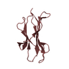
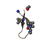
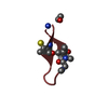
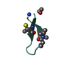
- PDB-3m17: Crystal structure of human FcRn with a monomeric peptide inhibitor -
+ Open data

- Basic information
| Entry | Database: PDB / ID: 3m17 | ||||||
|---|---|---|---|---|---|---|---|
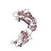
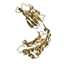
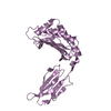
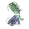
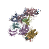
| Title | Crystal structure of human FcRn with a monomeric peptide inhibitor | ||||||
 Components Components |
| ||||||
 Keywords Keywords | IMMUNE SYSTEM/INHIBITOR / IMMUNOGLOBULIN BINDING PROTEIN / Cell membrane / Disulfide bond / Glycoprotein / IgG-binding protein / Immunoglobulin domain / Membrane / Receptor / Transmembrane / Amyloid / Amyloidosis / Disease mutation / Glycation / Immune response / MHC I / Pyrrolidone carboxylic acid / Secreted / IMMUNE SYSTEM-INHIBITOR complex | ||||||
| Function / homology |  Function and homology information Function and homology informationIgG immunoglobulin transcytosis in epithelial cells mediated by FcRn immunoglobulin receptor / IgG binding / beta-2-microglobulin binding / positive regulation of ferrous iron binding / positive regulation of transferrin receptor binding / positive regulation of receptor binding / early endosome lumen / Nef mediated downregulation of MHC class I complex cell surface expression / negative regulation of receptor binding / DAP12 interactions ...IgG immunoglobulin transcytosis in epithelial cells mediated by FcRn immunoglobulin receptor / IgG binding / beta-2-microglobulin binding / positive regulation of ferrous iron binding / positive regulation of transferrin receptor binding / positive regulation of receptor binding / early endosome lumen / Nef mediated downregulation of MHC class I complex cell surface expression / negative regulation of receptor binding / DAP12 interactions / cellular response to iron ion / Endosomal/Vacuolar pathway / Antigen Presentation: Folding, assembly and peptide loading of class I MHC / cellular response to iron(III) ion / antigen processing and presentation of exogenous protein antigen via MHC class Ib, TAP-dependent / negative regulation of forebrain neuron differentiation / regulation of erythrocyte differentiation / peptide antigen assembly with MHC class I protein complex / ER to Golgi transport vesicle membrane / regulation of iron ion transport / response to molecule of bacterial origin / MHC class I peptide loading complex / HFE-transferrin receptor complex / T cell mediated cytotoxicity / antigen processing and presentation of endogenous peptide antigen via MHC class I / positive regulation of T cell cytokine production / MHC class I protein complex / multicellular organismal-level iron ion homeostasis / negative regulation of neurogenesis / positive regulation of receptor-mediated endocytosis / peptide antigen assembly with MHC class II protein complex / MHC class II protein complex / cellular response to nicotine / positive regulation of T cell mediated cytotoxicity / specific granule lumen / recycling endosome membrane / phagocytic vesicle membrane / peptide antigen binding / positive regulation of cellular senescence / negative regulation of epithelial cell proliferation / antigen processing and presentation of exogenous peptide antigen via MHC class II / Immunoregulatory interactions between a Lymphoid and a non-Lymphoid cell / Interferon gamma signaling / positive regulation of immune response / Modulation by Mtb of host immune system / positive regulation of T cell activation / sensory perception of smell / negative regulation of neuron projection development / positive regulation of protein binding / tertiary granule lumen / DAP12 signaling / MHC class II protein complex binding / early endosome membrane / late endosome membrane / iron ion transport / ER-Phagosome pathway / T cell differentiation in thymus / protein refolding / protein homotetramerization / intracellular iron ion homeostasis / amyloid fibril formation / learning or memory / endosome membrane / immune response / Amyloid fiber formation / Golgi membrane / endoplasmic reticulum lumen / external side of plasma membrane / lysosomal membrane / focal adhesion / Neutrophil degranulation / SARS-CoV-2 activates/modulates innate and adaptive immune responses / structural molecule activity / Golgi apparatus / endoplasmic reticulum / protein homodimerization activity / extracellular space / extracellular exosome / extracellular region / identical protein binding / membrane / plasma membrane / cytosol Similarity search - Function | ||||||
| Biological species |  Homo sapiens (human) Homo sapiens (human) | ||||||
| Method |  X-RAY DIFFRACTION / SYNCHROTRON / MOLECULAR REPLACEMENT / Resolution: 2.6 Å X-RAY DIFFRACTION / SYNCHROTRON / MOLECULAR REPLACEMENT / Resolution: 2.6 Å | ||||||
 Authors Authors | Mezo, A.R. / Sridhar, V. / Badger, J. / Sakorafas, P. / Nienaber, V. | ||||||
 Citation Citation | Journal: J.Biol.Chem. / Year: 2010 Title: X-ray crystal structures of monomeric and dimeric peptide inhibitors in complex with the human neonatal Fc receptor, FcRn. Authors: Mezo, A.R. / Sridhar, V. / Badger, J. / Sakorafas, P. / Nienaber, V. | ||||||
| History |
|
- Structure visualization
| Structure viewer | Molecule: MolmilJmol/JSmol |
|---|
- Downloads & links
-Download
| PDBx/mmCIF format | 3m17.cif.gz | 292.3 KB | Display | PDBx/mmCIF format |
|---|---|---|---|---|
| PDB format | pdb3m17.ent.gz | 234.5 KB | Display | PDB format |
| PDBx/mmJSON format | 3m17.json.gz | Tree view | PDBx/mmJSON format | |
| Others |  Other downloads Other downloads |
-Validation report
| Summary document | 3m17_validation.pdf.gz | 509.4 KB | Display | wwPDB validaton report |
|---|---|---|---|---|
| Full document | 3m17_full_validation.pdf.gz | 525.8 KB | Display | |
| Data in XML | 3m17_validation.xml.gz | 52.6 KB | Display | |
| Data in CIF | 3m17_validation.cif.gz | 72.6 KB | Display | |
| Arichive directory | https://data.pdbj.org/pub/pdb/validation_reports/m1/3m17ftp://data.pdbj.org/pub/pdb/validation_reports/m1/3m17 | HTTPS FTP |
-Related structure data
| Related structure data |  3m1bC  1exuS C: citing same article ( S: Starting model for refinement |
|---|---|
| Similar structure data |


-Links
 PDBj
PDBj


- Assembly


-Components
| #1: Protein | Mass: 29720.383 Da / Num. of mol.: 4 / Fragment: UNP residues 24-290 Source method: isolated from a genetically manipulated source Source: (gene. exp.) Homo sapiens (human) / Gene: FCGRT, FCRN / Cell (production host): CHOK1SV cells / Production host:  Cricetinae (hamsters) / References: UniProt: P55899 Cricetinae (hamsters) / References: UniProt: P55899#2: Protein | Mass: 11748.160 Da / Num. of mol.: 4 / Fragment: UNP residues 21-119 Source method: isolated from a genetically manipulated source Source: (gene. exp.) Homo sapiens (human) / Gene: B2M, CDABP0092, HDCMA22P / Cell (production host): CHOK1SV cells / Production host: Cricetinae (hamsters) / References: UniProt: P61769#3: Protein/peptide | 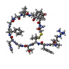  Type: Peptide-like / Class: Inhibitor / Mass: 1539.845 Da / Num. of mol.: 4 / Source method: obtained synthetically / Details: artificial peptide / References: monomeric peptide inhibitor Type: Peptide-like / Class: Inhibitor / Mass: 1539.845 Da / Num. of mol.: 4 / Source method: obtained synthetically / Details: artificial peptide / References: monomeric peptide inhibitor#4: Water | ChemComp-HOH / |  Mass: 18.015 Da / Num. of mol.: 183 / Source method: isolated from a natural source / Formula: H2O Mass: 18.015 Da / Num. of mol.: 183 / Source method: isolated from a natural source / Formula: H2O |
|---|
-Experimental details
-Experiment
| Experiment | Method: X-RAY DIFFRACTION / Number of used crystals: 1 |
|---|
- Sample preparation
| Crystal |
| |||||||||
|---|---|---|---|---|---|---|---|---|---|---|
| Crystal grow | Temperature: 277 K / Method: vapor diffusion, sitting drop / pH: 4.2 Details: 2uL protein:peptide with 2ul solution of 100mM sodium phosphate/citric acid, 20% PEG 3350, 8% ethanol, pH 4.2, VAPOR DIFFUSION, SITTING DROP, temperature 277K |
-Data collection
| Diffraction source | Source: SYNCHROTRON / Site: ALS  / Beamline: 4.2.2 / Wavelength: 1 Å / Beamline: 4.2.2 / Wavelength: 1 Å |
|---|---|
| Detector | Date: Sep 1, 2007 |
| Radiation | Protocol: SINGLE WAVELENGTH / Monochromatic (M) / Laue (L): M / Scattering type: x-ray |
| Radiation wavelength | Wavelength: 1 Å / Relative weight: 1 |
| Reflection | Resolution: 2.6→41.1 Å / Num. all: 51434 / Num. obs: 51434 / % possible obs: 98.7 % / Observed criterion σ(F): 0 / Observed criterion σ(I): -4 |
| Reflection shell | Resolution: 2.6→2.69 Å / % possible all: 97.8 |
- Processing
| Software |
| ||||||||||||||||||||||||||||||||||||||||||||||||||||||||||||||||||||||
|---|---|---|---|---|---|---|---|---|---|---|---|---|---|---|---|---|---|---|---|---|---|---|---|---|---|---|---|---|---|---|---|---|---|---|---|---|---|---|---|---|---|---|---|---|---|---|---|---|---|---|---|---|---|---|---|---|---|---|---|---|---|---|---|---|---|---|---|---|---|---|---|
| Refinement | Method to determine structure: MOLECULAR REPLACEMENT Starting model: PDB ENTRY 1EXU Resolution: 2.6→41.1 Å / Cor.coef. Fo:Fc: 0.894 / Cor.coef. Fo:Fc free: 0.827 / SU B: 15.11 / SU ML: 0.32 / Cross valid method: THROUGHOUT / ESU R: 1.51 / ESU R Free: 0.42 / Stereochemistry target values: MAXIMUM LIKELIHOOD
| ||||||||||||||||||||||||||||||||||||||||||||||||||||||||||||||||||||||
| Solvent computation | Ion probe radii: 0.8 Å / Shrinkage radii: 0.8 Å / VDW probe radii: 1.2 Å / Solvent model: MASK | ||||||||||||||||||||||||||||||||||||||||||||||||||||||||||||||||||||||
| Displacement parameters | Biso mean: 31.112 Å2
| ||||||||||||||||||||||||||||||||||||||||||||||||||||||||||||||||||||||
| Refinement step | Cycle: LAST / Resolution: 2.6→41.1 Å
| ||||||||||||||||||||||||||||||||||||||||||||||||||||||||||||||||||||||
| Refine LS restraints |
| ||||||||||||||||||||||||||||||||||||||||||||||||||||||||||||||||||||||
| LS refinement shell | Resolution: 2.6→2.667 Å / Total num. of bins used: 20
|
