 Movie
Movie Controller

[English] 日本語
 Yorodumi
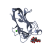
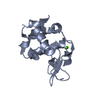
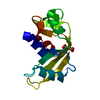
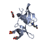
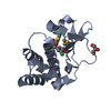
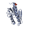
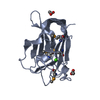
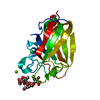
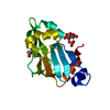
- PDB-3lgl: Crystal structure of the 53BP1 tandem tudor domain in complex wit... -
+ Open data

- Basic information
| Entry | Database: PDB / ID: 3lgl | ||||||
|---|---|---|---|---|---|---|---|
| Title | Crystal structure of the 53BP1 tandem tudor domain in complex with p53K382me2 | ||||||
 Components Components |
| ||||||
 Keywords Keywords |  CELL CYCLE / TANDEM TUDOR DOMAIN / DIMETHYLATED p53 PEPTIDE / DNA REPAIR / DNA damage / DNA-binding / Methylation / Transcription / Transcription regulation CELL CYCLE / TANDEM TUDOR DOMAIN / DIMETHYLATED p53 PEPTIDE / DNA REPAIR / DNA damage / DNA-binding / Methylation / Transcription / Transcription regulation | ||||||
| Function / homology |  Function and homology information Function and homology informationubiquitin-modified histone reader activity / positive regulation of isotype switching / cellular response to X-ray / double-strand break repair via classical nonhomologous end joining / DNA repair complex / negative regulation of double-strand break repair via homologous recombination / telomeric DNA binding / SUMOylation of transcription factors / methylated histone binding / histone reader activity ...ubiquitin-modified histone reader activity / positive regulation of isotype switching / cellular response to X-ray / double-strand break repair via classical nonhomologous end joining / DNA repair complex / negative regulation of double-strand break repair via homologous recombination / telomeric DNA binding / SUMOylation of transcription factors / methylated histone binding / histone reader activity / replication fork / DNA damage checkpoint signaling / transcription coregulator activity / Nonhomologous End-Joining (NHEJ) / protein homooligomerization / G2/M DNA damage checkpoint / kinetochore / double-strand break repair via nonhomologous end joining / positive regulation of DNA-binding transcription factor activity / p53 binding / Recruitment and ATM-mediated phosphorylation of repair and signaling proteins at DNA double strand breaks / site of double-strand break / Processing of DNA double-strand break ends / histone binding / RNA polymerase II-specific DNA-binding transcription factor binding / damaged DNA binding / chromosome, telomeric region / nuclear body / DNA damage response / positive regulation of DNA-templated transcription / positive regulation of transcription by RNA polymerase II / nucleoplasm / nucleus / cytoplasmSimilarity search - Function | ||||||
| Biological species |  Homo sapiens (human) Homo sapiens (human) | ||||||
| Method | X-RAY DIFFRACTION / SYNCHROTRON / MOLECULAR REPLACEMENT / Resolution: 1.6 Å | ||||||
 Authors Authors | Roy, S. / Kutateladze, T.G. | ||||||
 Citation Citation | Journal: J.Mol.Biol. / Year: 2010 Title: Structural insight into p53 recognition by the 53BP1 tandem Tudor domain. Authors: Roy, S. / Musselman, C.A. / Kachirskaia, I. / Hayashi, R. / Glass, K.C. / Nix, J.C. / Gozani, O. / Appella, E. / Kutateladze, T.G. | ||||||
| History |
|
- Structure visualization
| Structure viewer | Molecule: MolmilJmol/JSmol |
|---|
- Downloads & links
-Download
| PDBx/mmCIF format | 3lgl.cif.gz | 92.6 KB | Display | PDBx/mmCIF format |
|---|---|---|---|---|
| PDB format | pdb3lgl.ent.gz | 71.2 KB | Display | PDB format |
| PDBx/mmJSON format | 3lgl.json.gz | Tree view | PDBx/mmJSON format | |
| Others |  Other downloads Other downloads |
-Validation report
| Arichive directory | https://data.pdbj.org/pub/pdb/validation_reports/lg/3lglftp://data.pdbj.org/pub/pdb/validation_reports/lg/3lgl | HTTPS FTP |
|---|
-Related structure data
| Related structure data |  3lgfC 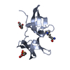 3lh0C  2g3rS C: citing same article ( S: Starting model for refinement |
|---|---|
| Similar structure data |
-Links
 PDBj
PDBj


- Assembly
| Deposited unit | 
| |||||||||||||||
|---|---|---|---|---|---|---|---|---|---|---|---|---|---|---|---|---|
| 1 |
| |||||||||||||||
| Unit cell |
| |||||||||||||||
| Components on special symmetry positions |
|
-Components
| #1: Protein | Mass: 14029.840 Da / Num. of mol.: 1 / Fragment: TANDEM TUDOR DOMAINS (RESIUDES 1484-1603) Source method: isolated from a genetically manipulated source Source: (gene. exp.) Homo sapiens (human) / Gene: TP53BP1 / Plasmid: pGEX-6P-1 / Production host:  Escherichia coli (E. coli) / Strain (production host): BL21(DE3) / References: UniProt: Q12888 Escherichia coli (E. coli) / Strain (production host): BL21(DE3) / References: UniProt: Q12888 |
|---|---|
| #2: Protein/peptide | Mass: 1408.754 Da / Num. of mol.: 1 / Source method: obtained synthetically / Details: The peptide was chemically synthesized |
| #3: Chemical | ChemComp-PGE / Polyethylene glycol  Mass: 150.173 Da / Num. of mol.: 1 / Source method: obtained synthetically / Formula: C6H14O4 Mass: 150.173 Da / Num. of mol.: 1 / Source method: obtained synthetically / Formula: C6H14O4 |
| #4: Chemical | ChemComp-SO4 / Sulfate  Mass: 96.063 Da / Num. of mol.: 1 / Source method: obtained synthetically / Formula: SO4 Mass: 96.063 Da / Num. of mol.: 1 / Source method: obtained synthetically / Formula: SO4 |
| #5: Water | ChemComp-HOH / Water Mass: 18.015 Da / Num. of mol.: 125 / Source method: isolated from a natural source / Formula: H2O Mass: 18.015 Da / Num. of mol.: 125 / Source method: isolated from a natural source / Formula: H2O |
-Experimental details
-Experiment
| Experiment | Method: X-RAY DIFFRACTION / Number of used crystals: 1 |
|---|
- Sample preparation
| Crystal | Density Matthews: 2.19 Å3/Da / Density % sol: 43.79 % |
|---|---|
| Crystal grow | Temperature: 298 K / Method: vapor diffusion, hanging drop / pH: 7 Details: 0.1 M HEPES-Na pH 7.0, 2% PEG 400 and 2.4 M ammonium sulphate, VAPOR DIFFUSION, HANGING DROP, temperature 298K |
-Data collection
| Diffraction | Mean temperature: 100 K |
|---|---|
| Diffraction source | Source: SYNCHROTRON / Site: ALS  / Beamline: 4.2.2 / Wavelength: 1 Å / Beamline: 4.2.2 / Wavelength: 1 Å |
| Detector | Type: NOIR-1 / Detector: CCD / Date: Jan 11, 2009 |
| Radiation | Monochromator: double crystal / Protocol: SINGLE WAVELENGTH / Monochromatic (M) / Laue (L): M / Scattering type: x-ray |
| Radiation wavelength | Wavelength: 1 Å / Relative weight: 1 |
| Reflection | Resolution: 1.6→39.03 Å / Num. all: 59066 / Num. obs: 59066 / % possible obs: 95.4 % / Observed criterion σ(F): 2 / Redundancy: 3.53 % / Biso Wilson estimate: 29.3 Å2 / Rmerge(I) obs: 0.053 / Net I/σ(I): 14.3 |
| Reflection shell | Resolution: 1.6→1.66 Å / Redundancy: 3.63 % / Rmerge(I) obs: 0.143 / Mean I/σ(I) obs: 6 / Num. unique all: 1754 / % possible all: 94.4 |
- Processing
| Software |
| |||||||||||||||||||||||||||||||||||||||||||||||||
|---|---|---|---|---|---|---|---|---|---|---|---|---|---|---|---|---|---|---|---|---|---|---|---|---|---|---|---|---|---|---|---|---|---|---|---|---|---|---|---|---|---|---|---|---|---|---|---|---|---|---|
| Refinement | Method to determine structure: MOLECULAR REPLACEMENT Starting model: PDB entry 2G3R Resolution: 1.6→24.133 Å / SU ML: 0.52 / Isotropic thermal model: Isotropic / Cross valid method: THROUGHOUT / σ(F): 1.34 / Stereochemistry target values: ML
| |||||||||||||||||||||||||||||||||||||||||||||||||
| Solvent computation | Shrinkage radii: 0.9 Å / VDW probe radii: 1.11 Å / Solvent model: FLAT BULK SOLVENT MODEL / Bsol: 55.826 Å2 / ksol: 0.451 e/Å3 | |||||||||||||||||||||||||||||||||||||||||||||||||
| Displacement parameters | Biso mean: 31.1 Å2
| |||||||||||||||||||||||||||||||||||||||||||||||||
| Refine analyze | Luzzati sigma a obs: 0.247 Å | |||||||||||||||||||||||||||||||||||||||||||||||||
| Refinement step | Cycle: LAST / Resolution: 1.6→24.133 Å
| |||||||||||||||||||||||||||||||||||||||||||||||||
| Refine LS restraints |
| |||||||||||||||||||||||||||||||||||||||||||||||||
| LS refinement shell |
|
