 Movie
Movie Controller

[English] 日本語
 Yorodumi
- PDB-3kov: Structure of MEF2A bound to DNA reveals a completely folded MADS-... -
+ Open data

- Basic information
| Entry | Database: PDB / ID: 3kov | ||||||
|---|---|---|---|---|---|---|---|
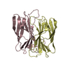
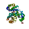
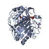
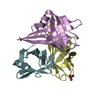
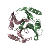
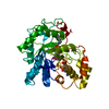
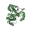
| Title | Structure of MEF2A bound to DNA reveals a completely folded MADS-box/MEF2 domain that recognizes DNA and recruits transcription co-factors | ||||||
 Components Components |
| ||||||
 Keywords Keywords | TRANSCRIPTION/DNA / MADS-box/MEF2 domain / transcription co-factors /  protein-DNA complex / protein-protein docking / Acetylation / Activator / Alternative splicing / Apoptosis / Developmental protein / Differentiation / Disease mutation / DNA-binding / Isopeptide bond / Neurogenesis / Nucleus / Phosphoprotein / Transcription / Transcription regulation / Ubl conjugation / TRANSCRIPTION-DNA complex protein-DNA complex / protein-protein docking / Acetylation / Activator / Alternative splicing / Apoptosis / Developmental protein / Differentiation / Disease mutation / DNA-binding / Isopeptide bond / Neurogenesis / Nucleus / Phosphoprotein / Transcription / Transcription regulation / Ubl conjugation / TRANSCRIPTION-DNA complex | ||||||
| Function / homology |  Function and homology information Function and homology informationERK5 cascade / ventricular cardiac myofibril assembly / mitochondrion distribution / cardiac conduction / mitochondrial genome maintenance / dendrite morphogenesis / muscle organ development / histone acetyltransferase binding / Myogenesis / positive regulation of cardiac muscle hypertrophy ...ERK5 cascade / ventricular cardiac myofibril assembly / mitochondrion distribution / cardiac conduction / mitochondrial genome maintenance / dendrite morphogenesis / muscle organ development / histone acetyltransferase binding / Myogenesis / positive regulation of cardiac muscle hypertrophy / SMAD binding / ERK/MAPK targets / cellular response to calcium ion / positive regulation of glucose import / RNA polymerase II transcription regulatory region sequence-specific DNA binding / histone deacetylase binding / MAPK cascade / heart development / DNA-binding transcription activator activity, RNA polymerase II-specific / DNA-binding transcription factor binding / transcription regulator complex / RNA polymerase II-specific DNA-binding transcription factor binding / sequence-specific DNA binding / cell differentiation / DNA-binding transcription factor activity, RNA polymerase II-specific / RNA polymerase II cis-regulatory region sequence-specific DNA binding / DNA-binding transcription factor activity / protein heterodimerization activity / DNA-templated transcription / apoptotic process / chromatin binding / chromatin / positive regulation of gene expression / protein kinase binding / negative regulation of transcription by RNA polymerase II / positive regulation of transcription by RNA polymerase II / nucleoplasm / nucleus / cytosolSimilarity search - Function | ||||||
| Biological species |  Homo sapiens (human) Homo sapiens (human) | ||||||
| Method | X-RAY DIFFRACTION / MOLECULAR REPLACEMENT / Resolution: 2.9 Å | ||||||
 Authors Authors | Wu, Y. / Dey, R. / Han, A. / Jayathilaka, N. / Philips, M. / Ye, J. / Chen, L. | ||||||
 Citation Citation | Journal: J.Mol.Biol. / Year: 2010 Title: Structure of the MADS-box/MEF2 Domain of MEF2A Bound to DNA and Its Implication for Myocardin Recruitment. Authors: Wu, Y. / Dey, R. / Han, A. / Jayathilaka, N. / Philips, M. / Ye, J. / Chen, L. | ||||||
| History |
|
- Structure visualization
| Structure viewer | Molecule: MolmilJmol/JSmol |
|---|
- Downloads & links
-Download
| PDBx/mmCIF format | 3kov.cif.gz | 238.3 KB | Display | PDBx/mmCIF format |
|---|---|---|---|---|
| PDB format | pdb3kov.ent.gz | 196.9 KB | Display | PDB format |
| PDBx/mmJSON format | 3kov.json.gz | Tree view | PDBx/mmJSON format | |
| Others |  Other downloads Other downloads |
-Validation report
| Arichive directory | https://data.pdbj.org/pub/pdb/validation_reports/ko/3kovftp://data.pdbj.org/pub/pdb/validation_reports/ko/3kov | HTTPS FTP |
|---|
-Related structure data
| Similar structure data |
|---|


-Links
 PDBj
PDBj


- Assembly
| Deposited unit | 
| |||||||||||||||||||||||||||||||||||||||||||||||||
|---|---|---|---|---|---|---|---|---|---|---|---|---|---|---|---|---|---|---|---|---|---|---|---|---|---|---|---|---|---|---|---|---|---|---|---|---|---|---|---|---|---|---|---|---|---|---|---|---|---|---|
| 1 | 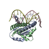
| |||||||||||||||||||||||||||||||||||||||||||||||||
| 2 | 
| |||||||||||||||||||||||||||||||||||||||||||||||||
| Unit cell |
| |||||||||||||||||||||||||||||||||||||||||||||||||
| Noncrystallographic symmetry (NCS) | NCS domain:
NCS domain segments:
NCS ensembles :
|
-Components
| #1: Protein | Mass: 10628.313 Da / Num. of mol.: 4 Source method: isolated from a genetically manipulated source Source: (gene. exp.) Homo sapiens (human) / Gene: MEF2A, MEF2 / Production host:  Escherichia coli (E. coli) / References: UniProt: Q02078 Escherichia coli (E. coli) / References: UniProt: Q02078#2: DNA chain | Mass: 3973.635 Da / Num. of mol.: 2 / Source method: obtained synthetically #3: DNA chain | Mass: 3964.621 Da / Num. of mol.: 2 / Source method: obtained synthetically |
|---|
-Experimental details
-Experiment
| Experiment | Method: X-RAY DIFFRACTION / Number of used crystals: 1 |
|---|
- Sample preparation
| Crystal | Density Matthews: 2.77 Å3/Da / Density % sol: 55.65 % | ||||||||||||||||||||||||||||||||||||||||||||||||||||
|---|---|---|---|---|---|---|---|---|---|---|---|---|---|---|---|---|---|---|---|---|---|---|---|---|---|---|---|---|---|---|---|---|---|---|---|---|---|---|---|---|---|---|---|---|---|---|---|---|---|---|---|---|---|
| Crystal grow | Temperature: 291 K / Method: hanging drop / pH: 4.7 Details: 50 mM acetic acid, 142mM NaCl, 5mM MgCl2, 10mM CaCl2, 3.3% Glycerol, 22.5% 3K PEG, pH 4.7, hanging drop, temperature 291K | ||||||||||||||||||||||||||||||||||||||||||||||||||||
| Components of the solutions |
|
-Data collection
| Diffraction source | Source: ROTATING ANODE / Type: OTHER / Wavelength: 1.5418 Å | ||||||||||||||||||||||||||||||||||||||||||||
|---|---|---|---|---|---|---|---|---|---|---|---|---|---|---|---|---|---|---|---|---|---|---|---|---|---|---|---|---|---|---|---|---|---|---|---|---|---|---|---|---|---|---|---|---|---|
| Detector | Type: RIGAKU RAXIS IV++ / Detector: IMAGE PLATE | ||||||||||||||||||||||||||||||||||||||||||||
| Radiation | Protocol: SINGLE WAVELENGTH / Monochromatic (M) / Laue (L): M / Scattering type: x-ray | ||||||||||||||||||||||||||||||||||||||||||||
| Radiation wavelength | Wavelength: 1.5418 Å / Relative weight: 1 | ||||||||||||||||||||||||||||||||||||||||||||
| Reflection | Resolution: 2.87→50 Å / Num. obs: 15478 / % possible obs: 99.6 % / Redundancy: 7 % / Rmerge(I) obs: 0.052 / Net I/σ(I): 30 | ||||||||||||||||||||||||||||||||||||||||||||
| Reflection shell |
|
- Processing
| Software |
| |||||||||||||||||||||||||||||||||||||||||||||||||
|---|---|---|---|---|---|---|---|---|---|---|---|---|---|---|---|---|---|---|---|---|---|---|---|---|---|---|---|---|---|---|---|---|---|---|---|---|---|---|---|---|---|---|---|---|---|---|---|---|---|---|
| Refinement | Method to determine structure: MOLECULAR REPLACEMENT / Resolution: 2.9→44.054 Å / Occupancy max: 1 / Occupancy min: 0.5 / SU ML: 1.37 / σ(F): 1.34 / Phase error: 27.37 / Stereochemistry target values: ML
| |||||||||||||||||||||||||||||||||||||||||||||||||
| Solvent computation | Shrinkage radii: 0.9 Å / VDW probe radii: 1.11 Å / Solvent model: FLAT BULK SOLVENT MODEL / Bsol: 69.484 Å2 / ksol: 0.306 e/Å3 | |||||||||||||||||||||||||||||||||||||||||||||||||
| Displacement parameters | Biso mean: 86.824 Å2
| |||||||||||||||||||||||||||||||||||||||||||||||||
| Refinement step | Cycle: LAST / Resolution: 2.9→44.054 Å
| |||||||||||||||||||||||||||||||||||||||||||||||||
| Refine LS restraints |
| |||||||||||||||||||||||||||||||||||||||||||||||||
| Refine LS restraints NCS |
| |||||||||||||||||||||||||||||||||||||||||||||||||
| LS refinement shell |
| |||||||||||||||||||||||||||||||||||||||||||||||||
| Refinement TLS params. | Method: refined / Origin x: 22.3433 Å / Origin y: 32.0434 Å / Origin z: 80.5156 Å
| |||||||||||||||||||||||||||||||||||||||||||||||||
| Refinement TLS group | Selection details: all |
