 Movie
Movie Controller

[English] 日本語
 Yorodumi
- PDB-5eu0: FIC domain of Bep1 from Bartonella rochalimae in complex with BiaA -
+ Open data

- Basic information
| Entry | Database: PDB / ID: 5eu0 | ||||||
|---|---|---|---|---|---|---|---|
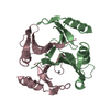
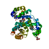
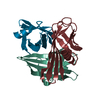
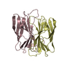
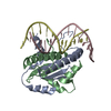
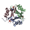
| Title | FIC domain of Bep1 from Bartonella rochalimae in complex with BiaA | ||||||
 Components Components |
| ||||||
 Keywords Keywords | TOXIN / AMPylation / adenylylation / Fic proteins / toxin-antitoxin complex | ||||||
| Function / homology |  Function and homology information Function and homology informationnegative regulation of protein adenylylation / protein adenylyltransferase / regulation of cell division / nucleotidyltransferase activity / ATP binding Similarity search - Function | ||||||
| Biological species |  Bartonella rochalimae ATCC BAA-1498 (bacteria) Bartonella rochalimae ATCC BAA-1498 (bacteria) | ||||||
| Method |  X-RAY DIFFRACTION / SYNCHROTRON / MOLECULAR REPLACEMENT / molecular replacement / Resolution: 1.6 Å X-RAY DIFFRACTION / SYNCHROTRON / MOLECULAR REPLACEMENT / molecular replacement / Resolution: 1.6 Å | ||||||
 Authors Authors | Goepfert, A. | ||||||
| Funding support |  Switzerland, 1items Switzerland, 1items
| ||||||
 Citation Citation | Journal: to be published Title: Molecular basis for Rho-family GTPase discrimination by a bacterial virulence factor Authors: Dietz, N. / Harms, A. / Sorg, I. / Mas, G. / Goepfert, A. / Hiller, S. / Schirmer, T. / Dehio, C. | ||||||
| History |
|
- Structure visualization
| Structure viewer | Molecule: MolmilJmol/JSmol |
|---|
- Downloads & links
-Download
| PDBx/mmCIF format | 5eu0.cif.gz | 132.2 KB | Display | PDBx/mmCIF format |
|---|---|---|---|---|
| PDB format | pdb5eu0.ent.gz | 103.7 KB | Display | PDB format |
| PDBx/mmJSON format | 5eu0.json.gz | Tree view | PDBx/mmJSON format | |
| Others |  Other downloads Other downloads |
-Validation report
| Arichive directory | https://data.pdbj.org/pub/pdb/validation_reports/eu/5eu0ftp://data.pdbj.org/pub/pdb/validation_reports/eu/5eu0 | HTTPS FTP |
|---|
-Related structure data
| Related structure data | 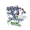 3shgS S: Starting model for refinement |
|---|---|
| Similar structure data |


-Links
 PDBj
PDBj- Assembly
| Deposited unit | 
| ||||||||
|---|---|---|---|---|---|---|---|---|---|
| 1 |
| ||||||||
| Unit cell |
|
-Components
| #1: Protein | Mass: 25997.557 Da / Num. of mol.: 1 / Fragment: FIC domain Source method: isolated from a genetically manipulated source Source: (gene. exp.) Bartonella rochalimae ATCC BAA-1498 (bacteria)Gene: BARRO_10061 / Plasmid: pRSFDuet1 / Production host: | ||
|---|---|---|---|
| #2: Protein | Mass: 7447.257 Da / Num. of mol.: 1 Source method: isolated from a genetically manipulated source Source: (gene. exp.) Bartonella rochalimae ATCC BAA-1498 (bacteria)Gene: BARRO_50056, O99_01280 / Plasmid: pRSFDuet1 / Production host: | ||
| #3: Chemical |   Mass: 96.063 Da / Num. of mol.: 2 / Source method: obtained synthetically / Formula: SO4 Mass: 96.063 Da / Num. of mol.: 2 / Source method: obtained synthetically / Formula: SO4#4: Water | ChemComp-HOH / |  Mass: 18.015 Da / Num. of mol.: 268 / Source method: isolated from a natural source / Formula: H2O Mass: 18.015 Da / Num. of mol.: 268 / Source method: isolated from a natural source / Formula: H2O |
-Experimental details
-Experiment
| Experiment | Method: X-RAY DIFFRACTION / Number of used crystals: 1 |
|---|
- Sample preparation
| Crystal | Density Matthews: 2.82 Å3/Da / Density % sol: 56.46 % |
|---|---|
| Crystal grow | Temperature: 277.15 K / Method: vapor diffusion, hanging drop / pH: 7.5 Details: 0.2M HEPES pH7.5, 2.3M AMMONIUM SULFATE, 2% v/v PEG 400 |
-Data collection
| Diffraction | Mean temperature: 100 K |
|---|---|
| Diffraction source | Source: SYNCHROTRON / Site: SLS / Beamline: X06DA / Wavelength: 1 Å |
| Detector | Type: MARMOSAIC 225 mm CCD / Detector: CCD / Date: Apr 14, 2011 |
| Radiation | Protocol: SINGLE WAVELENGTH / Monochromatic (M) / Laue (L): M / Scattering type: x-ray |
| Radiation wavelength | Wavelength: 1 Å / Relative weight: 1 |
| Reflection | Resolution: 1.6→73.1 Å / Num. obs: 47293 / % possible obs: 99.9 % / Observed criterion σ(I): -3 / Redundancy: 11.6 % / Biso Wilson estimate: 21 Å2 / CC1/2: 0.999 / Rmerge(I) obs: 0.14 / Rrim(I) all: 0.146 / Χ2: 1.013 / Net I/σ(I): 16.29 / Num. measured all: 547927 |
| Reflection shell | Resolution: 1.6→1.64 Å / Redundancy: 9.1 % / Rmerge(I) obs: 0.027 / Mean I/σ(I) obs: 1.31 / Num. measured obs: 6418 / Num. possible: 634 / Num. unique obs: 633 / CC1/2: 1 / Rrim(I) all: 0.028 / Rejects: 0 / % possible all: 98.5 |
-Phasing
| Phasing | Method: molecular replacement |
|---|
- Processing
| Software |
| ||||||||||||||||||||||||||||||||||||||||||||||||||||||||||||||||||||||||||||||||||||||||||||||||||||||||||||
|---|---|---|---|---|---|---|---|---|---|---|---|---|---|---|---|---|---|---|---|---|---|---|---|---|---|---|---|---|---|---|---|---|---|---|---|---|---|---|---|---|---|---|---|---|---|---|---|---|---|---|---|---|---|---|---|---|---|---|---|---|---|---|---|---|---|---|---|---|---|---|---|---|---|---|---|---|---|---|---|---|---|---|---|---|---|---|---|---|---|---|---|---|---|---|---|---|---|---|---|---|---|---|---|---|---|---|---|---|---|
| Refinement | Method to determine structure: MOLECULAR REPLACEMENT Starting model: 3SHG Resolution: 1.6→30 Å / Cor.coef. Fo:Fc: 0.955 / Cor.coef. Fo:Fc free: 0.948 / Rfactor Rfree error: 0 / SU R Cruickshank DPI: 0.077 / Cross valid method: THROUGHOUT / σ(F): 0 / SU R Blow DPI: 0.083 / SU Rfree Blow DPI: 0.08 / SU Rfree Cruickshank DPI: 0.077
| ||||||||||||||||||||||||||||||||||||||||||||||||||||||||||||||||||||||||||||||||||||||||||||||||||||||||||||
| Displacement parameters | Biso max: 97.71 Å2 / Biso mean: 25.19 Å2 / Biso min: 8.58 Å2
| ||||||||||||||||||||||||||||||||||||||||||||||||||||||||||||||||||||||||||||||||||||||||||||||||||||||||||||
| Refine analyze | Luzzati coordinate error obs: 0.22 Å | ||||||||||||||||||||||||||||||||||||||||||||||||||||||||||||||||||||||||||||||||||||||||||||||||||||||||||||
| Refinement step | Cycle: final / Resolution: 1.6→30 Å
| ||||||||||||||||||||||||||||||||||||||||||||||||||||||||||||||||||||||||||||||||||||||||||||||||||||||||||||
| Refine LS restraints |
| ||||||||||||||||||||||||||||||||||||||||||||||||||||||||||||||||||||||||||||||||||||||||||||||||||||||||||||
| LS refinement shell | Resolution: 1.6→1.64 Å / Total num. of bins used: 20
| ||||||||||||||||||||||||||||||||||||||||||||||||||||||||||||||||||||||||||||||||||||||||||||||||||||||||||||
| Refinement TLS params. | Method: refined / Refine-ID: X-RAY DIFFRACTION
| ||||||||||||||||||||||||||||||||||||||||||||||||||||||||||||||||||||||||||||||||||||||||||||||||||||||||||||
| Refinement TLS group |
|
