 Movie
Movie Controller

[English] 日本語
 Yorodumi
- PDB-3khn: Crystal structure of putative MotB like protein DVU_2228 from Des... -
+ Open data

- Basic information
| Entry | Database: PDB / ID: 3khn | ||||||
|---|---|---|---|---|---|---|---|
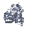
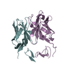
| Title | Crystal structure of putative MotB like protein DVU_2228 from Desulfovibrio vulgaris. | ||||||
 Components Components | MotB protein, putative | ||||||
 Keywords Keywords | structural genomics / unknown function / OmpA-like domain / PSI-2 / Protein Structure Initiative / New York SGX Research Center for Structural Genomics / NYSGXRC | ||||||
| Function / homology |  Function and homology information Function and homology information | ||||||
| Biological species |  Desulfovibrio vulgaris str. Hildenborough (bacteria) Desulfovibrio vulgaris str. Hildenborough (bacteria) | ||||||
| Method |  X-RAY DIFFRACTION / SYNCHROTRON / SAD / Resolution: 2.03 Å X-RAY DIFFRACTION / SYNCHROTRON / SAD / Resolution: 2.03 Å | ||||||
 Authors Authors | Ramagopal, U.A. / Toro, R. / Burley, S.K. / Almo, S.C. / New York SGX Research Center for Structural Genomics (NYSGXRC) | ||||||
 Citation Citation | Journal: To be Published Title: Crystal structure of putative MotB like protein DVU_2228 from Desulfovibrio vulgaris Authors: Ramagopal, U.A. / Toro, R. / Burley, S.K. / Almo, S.C. | ||||||
| History |
|
- Structure visualization
| Structure viewer | Molecule: MolmilJmol/JSmol |
|---|
- Downloads & links
-Download
| PDBx/mmCIF format | 3khn.cif.gz | 79 KB | Display | PDBx/mmCIF format |
|---|---|---|---|---|
| PDB format | pdb3khn.ent.gz | 59.7 KB | Display | PDB format |
| PDBx/mmJSON format | 3khn.json.gz | Tree view | PDBx/mmJSON format | |
| Others |  Other downloads Other downloads |
-Validation report
| Arichive directory | https://data.pdbj.org/pub/pdb/validation_reports/kh/3khnftp://data.pdbj.org/pub/pdb/validation_reports/kh/3khn | HTTPS FTP |
|---|
-Related structure data
| Similar structure data | |
|---|---|
| Other databases |
-Links
 PDBj
PDBj- Assembly
| Deposited unit | 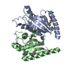
| ||||||||
|---|---|---|---|---|---|---|---|---|---|
| 1 |
| ||||||||
| Unit cell |
|
-Components
| #1: Protein | Mass: 19896.152 Da / Num. of mol.: 2 Source method: isolated from a genetically manipulated source Source: (gene. exp.) Desulfovibrio vulgaris str. Hildenborough (bacteria)Strain: Hildenborough / ATCC 29579 / NCIMB 8303 / Gene: DVU_2228 / Plasmid: pSGX4(BC) / Production host: #2: Water | ChemComp-HOH / |  Mass: 18.015 Da / Num. of mol.: 171 / Source method: isolated from a natural source / Formula: H2O Mass: 18.015 Da / Num. of mol.: 171 / Source method: isolated from a natural source / Formula: H2OHas protein modification | Y | |
|---|
-Experimental details
-Experiment
| Experiment | Method: X-RAY DIFFRACTION / Number of used crystals: 1 |
|---|
- Sample preparation
| Crystal | Density Matthews: 1.91 Å3/Da / Density % sol: 35.5 % |
|---|---|
| Crystal grow | Temperature: 298 K / Method: vapor diffusion, sitting drop / pH: 6.5 Details: 0.1M Bis-Tris pH 6.5, 30% PEG MME 550 0.05M Calcium Chloride, VAPOR DIFFUSION, SITTING DROP, temperature 298K |
-Data collection
| Diffraction | Mean temperature: 100 K |
|---|---|
| Diffraction source | Source: SYNCHROTRON / Site: NSLS  / Beamline: X29A / Wavelength: 0.9793 Å / Beamline: X29A / Wavelength: 0.9793 Å |
| Detector | Type: ADSC QUANTUM 315 / Detector: CCD / Date: Oct 5, 2009 |
| Radiation | Protocol: SINGLE WAVELENGTH / Scattering type: x-ray |
| Radiation wavelength | Wavelength: 0.9793 Å / Relative weight: 1 |
| Reflection | Resolution: 2.03→50 Å / Num. all: 20244 / Num. obs: 20244 / % possible obs: 98.9 % / Redundancy: 4.8 % / Rmerge(I) obs: 0.054 / Rsym value: 0.052 / Net I/σ(I): 30.3 |
| Reflection shell | Resolution: 2.03→2.07 Å / Redundancy: 4.5 % / Rmerge(I) obs: 0.086 / Mean I/σ(I) obs: 22.4 / Num. unique all: 793 / Rsym value: 0.09 / % possible all: 79.9 |
- Processing
| Software |
| |||||||||||||||||||||||||||||||||||||||||||||||||||||||||||||||||
|---|---|---|---|---|---|---|---|---|---|---|---|---|---|---|---|---|---|---|---|---|---|---|---|---|---|---|---|---|---|---|---|---|---|---|---|---|---|---|---|---|---|---|---|---|---|---|---|---|---|---|---|---|---|---|---|---|---|---|---|---|---|---|---|---|---|---|
| Refinement | Method to determine structure: SAD / Resolution: 2.03→50 Å / Cor.coef. Fo:Fc: 0.944 / Cor.coef. Fo:Fc free: 0.907 / WRfactor Rfree: 0.292 / WRfactor Rwork: 0.208 / Occupancy max: 1 / Occupancy min: 0.3 / FOM work R set: 0.835 / SU B: 4.374 / SU ML: 0.123 / SU R Cruickshank DPI: 0.22 / SU Rfree: 0.196 / Cross valid method: THROUGHOUT / σ(F): 0 / ESU R: 0.22 / ESU R Free: 0.196 / Stereochemistry target values: MAXIMUM LIKELIHOOD Details: HYDROGENS HAVE BEEN ADDED IN THE RIDING POSITIONS U VALUES : REFINED INDIVIDUALLY
| |||||||||||||||||||||||||||||||||||||||||||||||||||||||||||||||||
| Solvent computation | Ion probe radii: 0.8 Å / Shrinkage radii: 0.8 Å / VDW probe radii: 1.4 Å / Solvent model: MASK | |||||||||||||||||||||||||||||||||||||||||||||||||||||||||||||||||
| Displacement parameters | Biso max: 68.97 Å2 / Biso mean: 21.437 Å2 / Biso min: 6.21 Å2
| |||||||||||||||||||||||||||||||||||||||||||||||||||||||||||||||||
| Refinement step | Cycle: LAST / Resolution: 2.03→50 Å
| |||||||||||||||||||||||||||||||||||||||||||||||||||||||||||||||||
| Refine LS restraints |
| |||||||||||||||||||||||||||||||||||||||||||||||||||||||||||||||||
| LS refinement shell | Resolution: 2.033→2.086 Å / Total num. of bins used: 20
|
