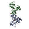
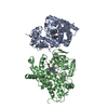
- PDB-3hrs: Crystal Structure of the Manganese-activated Repressor ScaR: apo form -
+
Open data
ID or keywords:
Loading...
-
Basic information
Entry
Database: PDB / ID: 3hrs
Title
Crystal Structure of the Manganese-activated Repressor ScaR: apo form
Components
Metalloregulator ScaR
Keywords
TRANSCRIPTION / DtxR/MntR family member
Function / homology
Function and homology information
transition metal ion binding / protein dimerization activity / DNA-binding transcription factor activity / DNA binding / cytoplasm Similarity search - Function
FeoA domain / Ferrous iron transport protein A (FeoA) / Ferrous iron transporter, core domain / Ferrous iron transporter FeoA domain / FeoA / : / DtxR-type HTH domain profile. / DTXR-type HTH domain / Iron dependent repressor, N-terminal DNA binding domain / Iron dependent repressor, metal binding and dimerisation domain ...FeoA domain / Ferrous iron transport protein A (FeoA) / Ferrous iron transporter, core domain / Ferrous iron transporter FeoA domain / FeoA / : / DtxR-type HTH domain profile. / DTXR-type HTH domain / Iron dependent repressor, N-terminal DNA binding domain / Iron dependent repressor, metal binding and dimerisation domain / Iron dependent repressor / Iron dependent repressor, metal binding and dimerisation domain superfamily / Iron dependent repressor, metal binding and dimerisation domain / Helix-turn-helix diphteria tox regulatory element / Transcriptional repressor, C-terminal / Winged helix-like DNA-binding domain superfamily/Winged helix DNA-binding domain / SH3 type barrels. / Arc Repressor Mutant, subunit A / Roll / Winged helix DNA-binding domain superfamily / Winged helix-like DNA-binding domain superfamily / Orthogonal Bundle / Mainly Beta / Mainly Alpha Similarity search - Domain/homology
Mass: 18.015 Da / Num. of mol.: 14 / Source method: isolated from a natural source / Formula: H2O
-
Experimental details
-
Experiment
Experiment
Method: X-RAY DIFFRACTION / Number of used crystals: 1
-
Sample preparation
Crystal
Density Matthews: 3.78 Å3/Da / Density % sol: 67.48 %
Crystal grow
Temperature: 298 K / Method: hanging drop / pH: 6 Details: 1.6 M lithium sulfate, 0.05 M sodium cacodylate in the presence of 1 mM MnCl2 and 1 mM duplex DNA containing operator sequence, pH 6.0, hanging drop, temperature 298K
-
Data collection
Diffraction
Mean temperature: 100 K
Diffraction source
Source: SYNCHROTRON / Site: ALS / Beamline: 8.2.1
Detector
Detector: CCD / Date: Jul 15, 2006
Radiation
Protocol: SINGLE WAVELENGTH / Monochromatic (M) / Laue (L): M / Scattering type: x-ray
Radiation wavelength
Relative weight: 1
Reflection
Resolution: 2.7→45.852 Å / Num. obs: 21511 / % possible obs: 97.5 % / Redundancy: 4.2 % / Rmerge(I) obs: 0.065 / Rsym value: 0.065 / Net I/σ(I): 7.674
Reflection shell
Resolution (Å)
Redundancy (%)
Rmerge(I) obs
Mean I/σ(I) obs
Num. measured all
Num. unique all
Rsym value
% possible all
2.7-2.85
3.5
0.317
2.4
9828
2771
0.317
90.7
2.85-3.02
4.1
0.229
3.2
11891
2880
0.229
98.2
3.02-3.23
4.6
0.162
4.4
12681
2782
0.162
99
3.23-3.49
4.5
0.111
6
11632
2572
0.111
99.5
3.49-3.82
4.5
0.088
6.9
10926
2414
0.088
99.2
3.82-4.27
4.4
0.068
8.9
9712
2196
0.068
99.5
4.27-4.93
4.4
0.057
10.7
8563
1968
0.057
99.6
4.93-6.04
4.3
0.052
11
7279
1683
0.052
99.2
6.04-8.54
4.2
0.043
12.8
5596
1344
0.043
99.1
8.54-45.85
3.4
0.038
16.4
2416
702
0.038
86.6
-
Processing
Software
Name
Version
Classification
NB
PHENIX
refinement
SCALA
3.1.20
datascaling
REFMAC
refinement
PDB_EXTRACT
3.005
dataextraction
ADSC
Quantum
datacollection
SOLVE
phasing
MOSFLM
datareduction
Refinement
Method to determine structure: SAD/SIR / Resolution: 2.7→20 Å / Occupancy max: 1 / Occupancy min: 1
In the structure databanks used in Yorodumi, some data are registered as the other names, "COVID-19 virus" and "2019-nCoV". Here are the details of the virus and the list of structure data.
Jan 31, 2019. EMDB accession codes are about to change! (news from PDBe EMDB page)
EMDB accession codes are about to change! (news from PDBe EMDB page)
The allocation of 4 digits for EMDB accession codes will soon come to an end. Whilst these codes will remain in use, new EMDB accession codes will include an additional digit and will expand incrementally as the available range of codes is exhausted. The current 4-digit format prefixed with “EMD-” (i.e. EMD-XXXX) will advance to a 5-digit format (i.e. EMD-XXXXX), and so on. It is currently estimated that the 4-digit codes will be depleted around Spring 2019, at which point the 5-digit format will come into force.
The EM Navigator/Yorodumi systems omit the EMD- prefix.
Related info.:Q: What is EMD? / ID/Accession-code notation in Yorodumi/EM Navigator
Yorodumi is a browser for structure data from EMDB, PDB, SASBDB, etc.
This page is also the successor to EM Navigator detail page, and also detail information page/front-end page for Omokage search.
The word "yorodu" (or yorozu) is an old Japanese word meaning "ten thousand". "mi" (miru) is to see.
Related info.:EMDB / PDB / SASBDB / Comparison of 3 databanks / Yorodumi Search / Aug 31, 2016. New EM Navigator & Yorodumi / Yorodumi Papers / Jmol/JSmol / Function and homology information / Changes in new EM Navigator and Yorodumi
 Movie
Movie Controller

 Yorodumi
 Open data
 Basic information
 Components
 Keywords
 Function and homology information
 Streptococcus gordonii (bacteria)
 X-RAY DIFFRACTION /
  Authors
 Citation
 Structure visualization
 Downloads & links
 Other downloads


 PDBj
PDBj Assembly

 Mass: 96.063 Da / Num. of mol.: 4 / Source method: obtained synthetically / Formula: SO4
 Mass: 18.015 Da / Num. of mol.: 14 / Source method: isolated from a natural source / Formula: H2O
 Sample preparation
 / Beamline: 8.2.1
 Processing