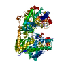
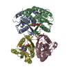
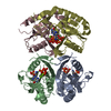
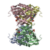
登録情報 データベース : PDB / ID : 3h83タイトル 2.06 Angstrom resolution structure of a hypoxanthine-guanine phosphoribosyltransferase (hpt-1) from Bacillus anthracis str. 'Ames Ancestor' Hypoxanthine phosphoribosyltransferase キーワード / / / / / / 機能・相同性 分子機能 ドメイン・相同性 構成要素
/ / / / / / / / / / / / / / / / / / / / / / / / / / / / / / / / 生物種 Bacillus anthracis str. 'Ames Ancestor' (炭疽菌)手法 / / / 解像度 : 2.06 Å データ登録者 Halavaty, A.S. / Shuvalova, L. / Minasov, G. / Dubrovska, I. / Peterson, S.N. / Anderson, W.F. / Center for Structural Genomics of Infectious Diseases (CSGID) ジャーナル : To be Published タイトル : 2.06 Angstrom resolution structure of a hypoxanthine-guanine phosphoribosyltransferase (hpt-1) from Bacillus anthracis str. 'Ames Ancestor'著者 : Halavaty, A.S. / Shuvalova, L. / Minasov, G. / Dubrovska, I. / Peterson, S.N. / Anderson, W.F. / Center for Structural Genomics of Infectious Diseases (CSGID) 履歴 登録 2009年4月28日 登録サイト / 処理サイト 改定 1.0 2009年5月12日 Provider / タイプ 改定 1.1 2011年7月13日 Group / Refinement description / Version format compliance改定 1.2 2017年11月1日 Group / カテゴリ / Item 改定 2.0 2020年7月29日 Group Advisory / Atomic model ... Advisory / Atomic model / Data collection / Database references / Derived calculations / Non-polymer description / Structure summary カテゴリ atom_site / chem_comp ... atom_site / chem_comp / database_PDB_caveat / entity / entity_name_com / pdbx_branch_scheme / pdbx_chem_comp_identifier / pdbx_entity_branch / pdbx_entity_branch_descriptor / pdbx_entity_branch_link / pdbx_entity_branch_list / pdbx_entity_nonpoly / pdbx_molecule_features / pdbx_nonpoly_scheme / pdbx_validate_chiral / struct_asym / struct_conn / struct_ref_seq_dif / struct_site / struct_site_gen Item _atom_site.B_iso_or_equiv / _atom_site.Cartn_x ... _atom_site.B_iso_or_equiv / _atom_site.Cartn_x / _atom_site.Cartn_y / _atom_site.Cartn_z / _atom_site.auth_asym_id / _atom_site.auth_atom_id / _atom_site.auth_comp_id / _atom_site.auth_seq_id / _atom_site.label_alt_id / _atom_site.label_asym_id / _atom_site.label_atom_id / _atom_site.label_comp_id / _atom_site.label_entity_id / _atom_site.occupancy / _atom_site.type_symbol / _chem_comp.formula / _chem_comp.formula_weight / _chem_comp.id / _chem_comp.mon_nstd_flag / _chem_comp.name / _chem_comp.type / _entity.formula_weight / _entity.pdbx_description / _entity.type / _pdbx_validate_chiral.auth_asym_id / _pdbx_validate_chiral.auth_comp_id / _pdbx_validate_chiral.auth_seq_id / _struct_asym.entity_id / _struct_ref_seq_dif.details 解説 / Provider / タイプ 改定 2.1 2023年9月6日 Group Data collection / Database references ... Data collection / Database references / Refinement description / Structure summary カテゴリ chem_comp / chem_comp_atom ... chem_comp / chem_comp_atom / chem_comp_bond / database_2 / pdbx_initial_refinement_model Item / _database_2.pdbx_DOI / _database_2.pdbx_database_accession
すべて表示 表示を減らす
 ムービー
ムービー コントローラー


 データを開く
 基本情報
基本情報 要素
要素 キーワード
キーワード 機能・相同性情報
 X線回折 /
  データ登録者
 引用
引用 構造の表示
構造の表示 ダウンロードとリンク
 その他のダウンロード
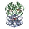

 PDBj
PDBj
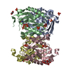
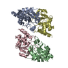
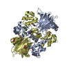
 集合体
集合体


 分子量: 94.971 Da / 分子数: 10 / 由来タイプ: 合成 / 式: PO4
 分子量: 18.015 Da / 分子数: 510 / 由来タイプ: 天然 / 式: H2O
 試料調製
試料調製 / ビームライン: 21-ID-G / 波長: 0.97856 Å
 解析
解析