 Movie
Movie Controller

[English] 日本語
 Yorodumi
- PDB-3t0c: Crystal structure of Streptococcus mutans MetE complexed with Zinc -
+ Open data

- Basic information
| Entry | Database: PDB / ID: 3t0c | |||||||||
|---|---|---|---|---|---|---|---|---|---|---|
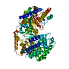
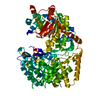
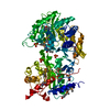
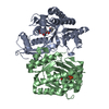
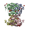
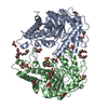
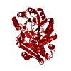
| Title | Crystal structure of Streptococcus mutans MetE complexed with Zinc | |||||||||
 Components Components | 5-methyltetrahydropteroyltriglutamate--homocysteine methyltransferase | |||||||||
 Keywords Keywords | TRANSFERASE / MetE / Barrel / methyltransferase | |||||||||
| Function / homology |  Function and homology information Function and homology information5-methyltetrahydropteroyltriglutamate-homocysteine S-methyltransferase / 5-methyltetrahydropteroyltriglutamate-homocysteine S-methyltransferase activity / 'de novo' L-methionine biosynthetic process / methylation / zinc ion binding Similarity search - Function | |||||||||
| Biological species |  Streptococcus mutans (bacteria) Streptococcus mutans (bacteria) | |||||||||
| Method |  X-RAY DIFFRACTION / MOLECULAR REPLACEMENT / Resolution: 2.187 Å X-RAY DIFFRACTION / MOLECULAR REPLACEMENT / Resolution: 2.187 Å | |||||||||
 Authors Authors | Fu, T.M. / Liang, Y.H. / Su, X.D. | |||||||||
 Citation Citation | Journal: J.Mol.Biol. / Year: 2011 Title: Crystal Structures of Cobalamin-Independent Methionine Synthase (MetE) from Streptococcus mutans: A Dynamic Zinc-Inversion Model Authors: Fu, T.M. / Almqvist, J. / Liang, Y.H. / Li, L. / Huang, Y. / Su, X.D. | |||||||||
| History |
|
- Structure visualization
| Structure viewer | Molecule: MolmilJmol/JSmol |
|---|
- Downloads & links
-Download
| PDBx/mmCIF format | 3t0c.cif.gz | 168.2 KB | Display | PDBx/mmCIF format |
|---|---|---|---|---|
| PDB format | pdb3t0c.ent.gz | 125.7 KB | Display | PDB format |
| PDBx/mmJSON format | 3t0c.json.gz | Tree view | PDBx/mmJSON format | |
| Others |  Other downloads Other downloads |
-Validation report
| Arichive directory | https://data.pdbj.org/pub/pdb/validation_reports/t0/3t0cftp://data.pdbj.org/pub/pdb/validation_reports/t0/3t0c | HTTPS FTP |
|---|
-Related structure data
| Related structure data |  3l7rC  2nq5S C: citing same article ( S: Starting model for refinement |
|---|---|
| Similar structure data |


-Links
 PDBj
PDBj- Assembly
| Deposited unit | 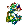
| ||||||||
|---|---|---|---|---|---|---|---|---|---|
| 1 |
| ||||||||
| Unit cell |
|
-Components
| #1: Protein | Mass: 87799.820 Da / Num. of mol.: 1 Source method: isolated from a genetically manipulated source Source: (gene. exp.) Streptococcus mutans (bacteria) / Strain: UA159 / Gene: metE / Plasmid: pET28a / Production host: References: UniProt: Q8CWX6, 5-methyltetrahydropteroyltriglutamate-homocysteine S-methyltransferase | ||||
|---|---|---|---|---|---|
| #2: Chemical |   Mass: 96.063 Da / Num. of mol.: 3 / Source method: obtained synthetically / Formula: SO4 Mass: 96.063 Da / Num. of mol.: 3 / Source method: obtained synthetically / Formula: SO4#3: Chemical | ChemComp-ZN / |   Mass: 65.409 Da / Num. of mol.: 1 / Source method: obtained synthetically / Formula: Zn Mass: 65.409 Da / Num. of mol.: 1 / Source method: obtained synthetically / Formula: Zn#4: Water | ChemComp-HOH / |  Mass: 18.015 Da / Num. of mol.: 493 / Source method: isolated from a natural source / Formula: H2O Mass: 18.015 Da / Num. of mol.: 493 / Source method: isolated from a natural source / Formula: H2O |
-Experimental details
-Experiment
| Experiment | Method: X-RAY DIFFRACTION / Number of used crystals: 1 |
|---|
- Sample preparation
| Crystal | Density Matthews: 2.32 Å3/Da / Density % sol: 47.08 % |
|---|---|
| Crystal grow | Temperature: 293 K / Method: vapor diffusion, hanging drop / pH: 7.5 Details: 0.2M Li2SO4, 0.1M HEPES, 25%(w/v) polyethylene glycerol 3350, pH 7.5, VAPOR DIFFUSION, HANGING DROP, temperature 293K |
-Data collection
| Diffraction | Mean temperature: 100 K |
|---|---|
| Diffraction source | Source: ROTATING ANODE / Type: BRUKER AXS MICROSTAR / Wavelength: 1.5418 Å |
| Detector | Type: BRUKER SMART 6000 / Detector: CCD / Date: May 8, 2005 |
| Radiation | Protocol: SINGLE WAVELENGTH / Monochromatic (M) / Laue (L): M / Scattering type: x-ray |
| Radiation wavelength | Wavelength: 1.5418 Å / Relative weight: 1 |
| Reflection | Resolution: 2.187→52.63 Å / Num. all: 40832 / Num. obs: 39151 / % possible obs: 95.88 % / Observed criterion σ(F): 3 / Observed criterion σ(I): 3 / Redundancy: 3.31 % / Biso Wilson estimate: 20.74 Å2 |
| Reflection shell | Resolution: 2.19→2.33 Å / Num. unique all: 6372 / % possible all: 77.2 |
- Processing
| Software |
| |||||||||||||||||||||||||||||||||||||||||||||||||||||||||||||||||||||||||||||||||||||||||||||||||||||||||
|---|---|---|---|---|---|---|---|---|---|---|---|---|---|---|---|---|---|---|---|---|---|---|---|---|---|---|---|---|---|---|---|---|---|---|---|---|---|---|---|---|---|---|---|---|---|---|---|---|---|---|---|---|---|---|---|---|---|---|---|---|---|---|---|---|---|---|---|---|---|---|---|---|---|---|---|---|---|---|---|---|---|---|---|---|---|---|---|---|---|---|---|---|---|---|---|---|---|---|---|---|---|---|---|---|---|---|
| Refinement | Method to determine structure: MOLECULAR REPLACEMENT Starting model: 2NQ5 Resolution: 2.187→42.068 Å / Occupancy max: 1 / Occupancy min: 0.18 / FOM work R set: 0.7848 / SU ML: 0.35 / σ(F): 1.91 / Phase error: 28.48 / Stereochemistry target values: ML
| |||||||||||||||||||||||||||||||||||||||||||||||||||||||||||||||||||||||||||||||||||||||||||||||||||||||||
| Solvent computation | Shrinkage radii: 0.9 Å / VDW probe radii: 1.11 Å / Solvent model: FLAT BULK SOLVENT MODEL / Bsol: 38.393 Å2 / ksol: 0.3 e/Å3 | |||||||||||||||||||||||||||||||||||||||||||||||||||||||||||||||||||||||||||||||||||||||||||||||||||||||||
| Displacement parameters | Biso max: 70.47 Å2 / Biso mean: 26.1002 Å2 / Biso min: 9.06 Å2
| |||||||||||||||||||||||||||||||||||||||||||||||||||||||||||||||||||||||||||||||||||||||||||||||||||||||||
| Refinement step | Cycle: LAST / Resolution: 2.187→42.068 Å
| |||||||||||||||||||||||||||||||||||||||||||||||||||||||||||||||||||||||||||||||||||||||||||||||||||||||||
| Refine LS restraints |
| |||||||||||||||||||||||||||||||||||||||||||||||||||||||||||||||||||||||||||||||||||||||||||||||||||||||||
| LS refinement shell | Refine-ID: X-RAY DIFFRACTION / Total num. of bins used: 14
|
