 Movie
Movie Controller


+ Open data

- Basic information
| Entry | Database: PDB / ID: 3geb | ||||||
|---|---|---|---|---|---|---|---|
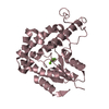
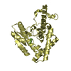
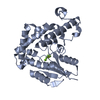
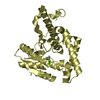
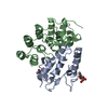
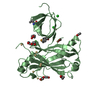
| Title | Crystal Structure of edeya2 | ||||||
 Components Components | Eyes absent homolog 2 | ||||||
 Keywords Keywords | HYDROLASE / Activator / Alternative splicing / Cytoplasm / Developmental protein / Magnesium / Nucleus / Polymorphism / Protein phosphatase / Transcription / Transcription regulation | ||||||
| Function / homology |  Function and homology information Function and homology informationhistone H2AXY142 phosphatase activity / mesodermal cell fate specification / striated muscle tissue development / mitochondrial outer membrane permeabilization / negative regulation of extrinsic apoptotic signaling pathway in absence of ligand / positive regulation of DNA repair / extrinsic apoptotic signaling pathway in absence of ligand / protein-tyrosine-phosphatase / protein tyrosine phosphatase activity / Recruitment and ATM-mediated phosphorylation of repair and signaling proteins at DNA double strand breaks ...histone H2AXY142 phosphatase activity / mesodermal cell fate specification / striated muscle tissue development / mitochondrial outer membrane permeabilization / negative regulation of extrinsic apoptotic signaling pathway in absence of ligand / positive regulation of DNA repair / extrinsic apoptotic signaling pathway in absence of ligand / protein-tyrosine-phosphatase / protein tyrosine phosphatase activity / Recruitment and ATM-mediated phosphorylation of repair and signaling proteins at DNA double strand breaks / cell differentiation / DNA repair / magnesium ion binding / mitochondrion / nucleoplasm / nucleus / cytosol Similarity search - Function | ||||||
| Biological species |  Homo sapiens (human) Homo sapiens (human) | ||||||
| Method |  X-RAY DIFFRACTION / SYNCHROTRON / MOLECULAR REPLACEMENT / Resolution: 2.4 Å X-RAY DIFFRACTION / SYNCHROTRON / MOLECULAR REPLACEMENT / Resolution: 2.4 Å | ||||||
 Authors Authors | Kim, S.J. | ||||||
 Citation Citation | Journal: To be Published Title: Structure of edeya2 Authors: Kim, S.J. / Jeong, D.G. / Jung, S.K. / Seong, E.R. | ||||||
| History |
|
- Structure visualization
| Structure viewer | Molecule: MolmilJmol/JSmol |
|---|
- Downloads & links
-Download
| PDBx/mmCIF format | 3geb.cif.gz | 209.6 KB | Display | PDBx/mmCIF format |
|---|---|---|---|---|
| PDB format | pdb3geb.ent.gz | 170.6 KB | Display | PDB format |
| PDBx/mmJSON format | 3geb.json.gz | Tree view | PDBx/mmJSON format | |
| Others |  Other downloads Other downloads |
-Validation report
| Summary document | 3geb_validation.pdf.gz | 463 KB | Display | wwPDB validaton report |
|---|---|---|---|---|
| Full document | 3geb_full_validation.pdf.gz | 497.4 KB | Display | |
| Data in XML | 3geb_validation.xml.gz | 40.3 KB | Display | |
| Data in CIF | 3geb_validation.cif.gz | 55 KB | Display | |
| Arichive directory | https://data.pdbj.org/pub/pdb/validation_reports/ge/3gebftp://data.pdbj.org/pub/pdb/validation_reports/ge/3geb | HTTPS FTP |
-Related structure data
| Similar structure data |
|---|


-Links
 PDBj
PDBj


- Assembly
| Deposited unit | 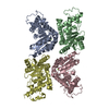
| ||||||||
|---|---|---|---|---|---|---|---|---|---|
| 1 | 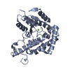
| ||||||||
| 2 | 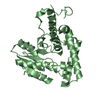
| ||||||||
| 3 | 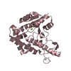
| ||||||||
| 4 | 
| ||||||||
| Unit cell |
|
-Components
| #1: Protein | Mass: 31054.330 Da / Num. of mol.: 4 / Fragment: C-terminal domain Source method: isolated from a genetically manipulated source Source: (gene. exp.) Homo sapiens (human) / Plasmid: pET21a / Production host:  #2: Chemical | ChemComp-MG /   Mass: 24.305 Da / Num. of mol.: 4 / Source method: obtained synthetically / Formula: Mg Mass: 24.305 Da / Num. of mol.: 4 / Source method: obtained synthetically / Formula: Mg#3: Water | ChemComp-HOH / |  Mass: 18.015 Da / Num. of mol.: 82 / Source method: isolated from a natural source / Formula: H2O Mass: 18.015 Da / Num. of mol.: 82 / Source method: isolated from a natural source / Formula: H2O |
|---|
-Experimental details
-Experiment
| Experiment | Method: X-RAY DIFFRACTION / Number of used crystals: 1 |
|---|
- Sample preparation
| Crystal | Density Matthews: 4.08 Å3/Da / Density % sol: 69.82 % |
|---|---|
| Crystal grow | Temperature: 291 K / Method: vapor diffusion, hanging drop / pH: 6 Details: Sodium chloride, pH 6.0, VAPOR DIFFUSION, HANGING DROP, temperature 291K |
-Data collection
| Diffraction | Mean temperature: 100 K | |||||||||||||||
|---|---|---|---|---|---|---|---|---|---|---|---|---|---|---|---|---|
| Diffraction source | Source: SYNCHROTRON / Site: PAL/PLS  / Beamline: 4A / Wavelength: 1 Å / Beamline: 4A / Wavelength: 1 Å | |||||||||||||||
| Detector | Type: ADSC QUANTUM 315 / Detector: CCD / Date: Jun 15, 2006 / Details: mirrors | |||||||||||||||
| Radiation | Monochromator: Mirrors / Protocol: SINGLE WAVELENGTH / Monochromatic (M) / Laue (L): M / Scattering type: x-ray | |||||||||||||||
| Radiation wavelength | Wavelength: 1 Å / Relative weight: 1 | |||||||||||||||
| Reflection twin |
| |||||||||||||||
| Reflection | Resolution: 2.4→50 Å / Num. all: 77791 / Num. obs: 76896 / % possible obs: 99.8 % / Observed criterion σ(F): 0 / Observed criterion σ(I): 0 / Redundancy: 4.6 % / Rmerge(I) obs: 0.079 / Rsym value: 0.079 / Net I/σ(I): 6.9 | |||||||||||||||
| Reflection shell | Resolution: 2.4→2.53 Å / Redundancy: 4.7 % / Rmerge(I) obs: 0.327 / Mean I/σ(I) obs: 2.5 / Num. unique all: 11332 / Rsym value: 0.293 / % possible all: 99.9 |
- Processing
| Software |
| |||||||||||||||||||||||||
|---|---|---|---|---|---|---|---|---|---|---|---|---|---|---|---|---|---|---|---|---|---|---|---|---|---|---|
| Refinement | Method to determine structure: MOLECULAR REPLACEMENT / Resolution: 2.4→50 Å / Isotropic thermal model: Isotropic / Cross valid method: THROUGHOUT / σ(F): 0 / σ(I): 0 / Stereochemistry target values: Engh & Huber Details: The crystal form is twinned by the operator h,-k,-l, twinning fraction 0.35
| |||||||||||||||||||||||||
| Displacement parameters | Biso mean: 41.7 Å2 | |||||||||||||||||||||||||
| Refinement step | Cycle: LAST / Resolution: 2.4→50 Å
|
