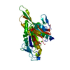
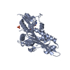
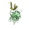
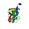
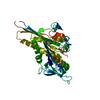
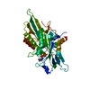
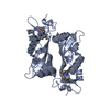
- PDB-3gbj: Crystal structure of the motor domain of kinesin KIF13B bound with ADP -
+
Open data
ID or keywords:
Loading...
-
Basic information
Entry
Database: PDB / ID: 3gbj
Title
Crystal structure of the motor domain of kinesin KIF13B bound with ADP
Components
KIF13B protein
Keywords
MOTOR PROTEIN / kinesin / motor domain / ADP / STRUCTURAL GENOMICS / STRUCTURAL GENOMICS CONSORTIUM / SGC / ATP-binding / Microtubule / Nucleotide-binding
Function / homology
Function and homology information
plus-end-directed vesicle transport along microtubule / cytoskeleton-dependent intracellular transport / Kinesins / COPI-dependent Golgi-to-ER retrograde traffic / microtubule motor activity / kinesin complex / regulation of axonogenesis / microtubule-based movement / protein targeting / 14-3-3 protein binding ...plus-end-directed vesicle transport along microtubule / cytoskeleton-dependent intracellular transport / Kinesins / COPI-dependent Golgi-to-ER retrograde traffic / microtubule motor activity / kinesin complex / regulation of axonogenesis / microtubule-based movement / protein targeting / 14-3-3 protein binding / T cell activation / microtubule binding / microtubule / axon / protein kinase binding / signal transduction / ATP hydrolysis activity / ATP binding / cytoplasm Similarity search - Function
Kinesin-like protein KIF13A / Kinesin-like KIF1-type / Kinesin protein 1B / Kinesin-like / Kinesin protein / CAP-Gly domain signature. / CAP Gly-rich domain / CAP Gly-rich domain superfamily / CAP-Gly domain / CAP-Gly domain profile. ...Kinesin-like protein KIF13A / Kinesin-like KIF1-type / Kinesin protein 1B / Kinesin-like / Kinesin protein / CAP-Gly domain signature. / CAP Gly-rich domain / CAP Gly-rich domain superfamily / CAP-Gly domain / CAP-Gly domain profile. / CAP_GLY / Kinesin-associated / Kinesin-associated / Kinesin motor domain / Kinesin / Kinesin-like protein / FHA domain / Forkhead-associated (FHA) domain / Kinesin motor domain signature. / Kinesin motor domain, conserved site / Kinesin motor domain / Kinesin motor domain profile. / Kinesin motor, catalytic domain. ATPase. / Kinesin motor domain / SMAD/FHA domain superfamily / Kinesin motor domain superfamily / P-loop containing nucleoside triphosphate hydrolase / 3-Layer(aba) Sandwich / Alpha Beta Similarity search - Domain/homology
Resolution: 2.102→39.062 Å / FOM work R set: 0.797 / Stereochemistry target values: MAXIMUM LIKELIHOOD Details: The Friedel pairs were used in phasing. Programs coot, molprobity have also been used in refinement
Rfactor
Num. reflection
% reflection
Selection details
Rfree
0.269
3474
2.67 %
Thin shells
Rwork
0.221
-
-
-
obs
0.222
133690
98.93 %
-
Solvent computation
Shrinkage radii: 0.9 Å / Solvent model: FLAT BULK SOLVENT MODEL / Bsol: 49.55 Å2 / ksol: 0.337 e/Å3
In the structure databanks used in Yorodumi, some data are registered as the other names, "COVID-19 virus" and "2019-nCoV". Here are the details of the virus and the list of structure data.
Jan 31, 2019. EMDB accession codes are about to change! (news from PDBe EMDB page)
EMDB accession codes are about to change! (news from PDBe EMDB page)
The allocation of 4 digits for EMDB accession codes will soon come to an end. Whilst these codes will remain in use, new EMDB accession codes will include an additional digit and will expand incrementally as the available range of codes is exhausted. The current 4-digit format prefixed with “EMD-” (i.e. EMD-XXXX) will advance to a 5-digit format (i.e. EMD-XXXXX), and so on. It is currently estimated that the 4-digit codes will be depleted around Spring 2019, at which point the 5-digit format will come into force.
The EM Navigator/Yorodumi systems omit the EMD- prefix.
Related info.:Q: What is EMD? / ID/Accession-code notation in Yorodumi/EM Navigator
Yorodumi is a browser for structure data from EMDB, PDB, SASBDB, etc.
This page is also the successor to EM Navigator detail page, and also detail information page/front-end page for Omokage search.
The word "yorodu" (or yorozu) is an old Japanese word meaning "ten thousand". "mi" (miru) is to see.
Related info.:EMDB / PDB / SASBDB / Comparison of 3 databanks / Yorodumi Search / Aug 31, 2016. New EM Navigator & Yorodumi / Yorodumi Papers / Jmol/JSmol / Function and homology information / Changes in new EM Navigator and Yorodumi
 Movie
Movie Controller

 Yorodumi
 Open data
 Basic information
 Components
 Keywords
 Function and homology information
 Homo sapiens (human)
 X-RAY DIFFRACTION /
  Authors
 Citation
 Structure visualization
 Downloads & links
 Other downloads
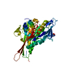

 PDBj
PDBj


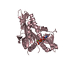
 Assembly
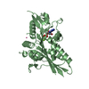


 Mass: 427.201 Da / Num. of mol.: 3 / Source method: obtained synthetically / Formula: C10H15N5O10P2 / Comment: ADP, energy-carrying molecule*YM
 Mass: 24.305 Da / Num. of mol.: 3 / Source method: obtained synthetically / Formula: Mg
 Num. of mol.: 5 / Source method: obtained synthetically
 Mass: 18.015 Da / Num. of mol.: 136 / Source method: isolated from a natural source / Formula: H2O
 Sample preparation
 / Beamline: 19-ID / Wavelength: 0.97937 Å
 Processing