 Movie
Movie Controller

[English] 日本語
 Yorodumi
- PDB-3f19: Crystal structure of the catalytic domain of human MMP12 complexe... -
+ Open data

- Basic information
| Entry | Database: PDB / ID: 3f19 | ||||||
|---|---|---|---|---|---|---|---|
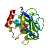
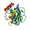
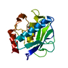
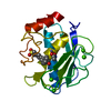
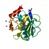
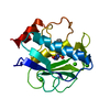
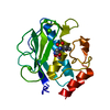
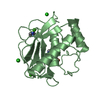
| Title | Crystal structure of the catalytic domain of human MMP12 complexed with the inhibitor 4-fluoro-N-(2-nitroso-2-oxoethyl)benzenesulfonamide | ||||||
 Components Components | Macrophage metalloelastase | ||||||
 Keywords Keywords |  HYDROLASE / MATRIX METALLOPROTEINASE / MMP12 / ELASTASE / COMPLEX (ELASTASE-INHIBITOR) / METALLO ELASTASE / CALCIUM / EXTRACELLULAR MATRIX / GLYCOPROTEIN / METAL-BINDING / METALLOPROTEASE / POLYMORPHISM / PROTEASE / SECRETED / ZINC / Zymogen HYDROLASE / MATRIX METALLOPROTEINASE / MMP12 / ELASTASE / COMPLEX (ELASTASE-INHIBITOR) / METALLO ELASTASE / CALCIUM / EXTRACELLULAR MATRIX / GLYCOPROTEIN / METAL-BINDING / METALLOPROTEASE / POLYMORPHISM / PROTEASE / SECRETED / ZINC / Zymogen | ||||||
| Function / homology |  Function and homology informationmacrophage elastase / negative regulation of endothelial cell-matrix adhesion via fibronectin / bronchiole development / positive regulation of epithelial cell proliferation involved in wound healing / elastin catabolic process / regulation of defense response to virus by host / positive regulation of type I interferon-mediated signaling pathway / wound healing, spreading of epidermal cells / negative regulation of type I interferon-mediated signaling pathway / lung alveolus development ...macrophage elastase / negative regulation of endothelial cell-matrix adhesion via fibronectin / bronchiole development / positive regulation of epithelial cell proliferation involved in wound healing / elastin catabolic process / regulation of defense response to virus by host / positive regulation of type I interferon-mediated signaling pathway / wound healing, spreading of epidermal cells / negative regulation of type I interferon-mediated signaling pathway / lung alveolus development / positive regulation of interferon-alpha production / response to amyloid-beta / Collagen degradation / collagen catabolic process / extracellular matrix disassembly / core promoter sequence-specific DNA binding / collagen binding / extracellular matrix / Degradation of the extracellular matrix / extracellular matrix organization / metalloendopeptidase activity / cellular response to virus / protein import into nucleus / endopeptidase activity / sequence-specific DNA binding / serine-type endopeptidase activity / calcium ion binding / negative regulation of transcription by RNA polymerase II / positive regulation of transcription by RNA polymerase II / proteolysis / extracellular space / zinc ion binding / extracellular region / nucleus / cytoplasm Function and homology informationmacrophage elastase / negative regulation of endothelial cell-matrix adhesion via fibronectin / bronchiole development / positive regulation of epithelial cell proliferation involved in wound healing / elastin catabolic process / regulation of defense response to virus by host / positive regulation of type I interferon-mediated signaling pathway / wound healing, spreading of epidermal cells / negative regulation of type I interferon-mediated signaling pathway / lung alveolus development ...macrophage elastase / negative regulation of endothelial cell-matrix adhesion via fibronectin / bronchiole development / positive regulation of epithelial cell proliferation involved in wound healing / elastin catabolic process / regulation of defense response to virus by host / positive regulation of type I interferon-mediated signaling pathway / wound healing, spreading of epidermal cells / negative regulation of type I interferon-mediated signaling pathway / lung alveolus development / positive regulation of interferon-alpha production / response to amyloid-beta / Collagen degradation / collagen catabolic process / extracellular matrix disassembly / core promoter sequence-specific DNA binding / collagen binding / extracellular matrix / Degradation of the extracellular matrix / extracellular matrix organization / metalloendopeptidase activity / cellular response to virus / protein import into nucleus / endopeptidase activity / sequence-specific DNA binding / serine-type endopeptidase activity / calcium ion binding / negative regulation of transcription by RNA polymerase II / positive regulation of transcription by RNA polymerase II / proteolysis / extracellular space / zinc ion binding / extracellular region / nucleus / cytoplasmSimilarity search - Function | ||||||
| Biological species |  Homo sapiens (human) Homo sapiens (human) | ||||||
| Method | X-RAY DIFFRACTION / SYNCHROTRON / MOLECULAR REPLACEMENT / Resolution: 1.13 Å | ||||||
 Authors Authors | Calderone, V. | ||||||
 Citation Citation | Journal: J.Am.Chem.Soc. / Year: 2007 Title: Exploring the subtleties of drug-receptor interactions: the case of matrix metalloproteinases. Authors: Bertini, I. / Calderone, V. / Fragai, M. / Giachetti, A. / Loconte, M. / Luchinat, C. / Maletta, M. / Nativi, C. / Yeo, K.J. #1: Journal: Angew.Chem.Int.Ed.Engl. / Year: 2003Title: X-ray structures of binary and ternary enzyme-product-inhibitor complexes of matrix metalloproteinases. Authors: Bertini, I. / Calderone, V. / Fragai, M. / Luchinat, C. / Mangani, S. / Terni, B. | ||||||
| History |
|
- Structure visualization
| Structure viewer | Molecule: MolmilJmol/JSmol |
|---|
- Downloads & links
-Download
| PDBx/mmCIF format | 3f19.cif.gz | 88.8 KB | Display | PDBx/mmCIF format |
|---|---|---|---|---|
| PDB format | pdb3f19.ent.gz | 66.5 KB | Display | PDB format |
| PDBx/mmJSON format | 3f19.json.gz | Tree view | PDBx/mmJSON format | |
| Others |  Other downloads Other downloads |
-Validation report
| Arichive directory | https://data.pdbj.org/pub/pdb/validation_reports/f1/3f19ftp://data.pdbj.org/pub/pdb/validation_reports/f1/3f19 | HTTPS FTP |
|---|
-Related structure data
| Related structure data |  3f15C  3f16C  3f17C  3f18C  3f1aC  3lk8C  3nx7C C: citing same article ( |
|---|---|
| Similar structure data |
-Links
 PDBj
PDBj


- Assembly
| Deposited unit | 
| ||||||||||||||||||
|---|---|---|---|---|---|---|---|---|---|---|---|---|---|---|---|---|---|---|---|
| 1 |
| ||||||||||||||||||
| Unit cell |
| ||||||||||||||||||
| Components on special symmetry positions |
|
-Components
| #1: Protein | Mass: 17484.475 Da / Num. of mol.: 1 / Fragment: CATALYTIC DOMAIN, UNP RESIDUES 106-263 / Mutation: F171D Source method: isolated from a genetically manipulated source Source: (gene. exp.) Homo sapiens (human) / Gene: HME, MMP12 / Plasmid: PET 21 / Production host:  Escherichia coli (E. coli) / Strain (production host): BL21 (DE3) / References: UniProt: P39900 Escherichia coli (E. coli) / Strain (production host): BL21 (DE3) / References: UniProt: P39900 | ||||||
|---|---|---|---|---|---|---|---|
| #2: Chemical |   Mass: 65.409 Da / Num. of mol.: 2 / Source method: obtained synthetically / Formula: Zn Mass: 65.409 Da / Num. of mol.: 2 / Source method: obtained synthetically / Formula: Zn#3: Chemical |   Mass: 40.078 Da / Num. of mol.: 3 / Source method: obtained synthetically / Formula: Ca Mass: 40.078 Da / Num. of mol.: 3 / Source method: obtained synthetically / Formula: Ca#4: Chemical |   Mass: 246.216 Da / Num. of mol.: 1 / Source method: obtained synthetically / Formula: C8H7FN2O4S Mass: 246.216 Da / Num. of mol.: 1 / Source method: obtained synthetically / Formula: C8H7FN2O4S#5: Water | ChemComp-HOH / | Water Mass: 18.015 Da / Num. of mol.: 323 / Source method: isolated from a natural source / Formula: H2O Mass: 18.015 Da / Num. of mol.: 323 / Source method: isolated from a natural source / Formula: H2O |
-Experimental details
-Experiment
| Experiment | Method: X-RAY DIFFRACTION / Number of used crystals: 1 |
|---|
- Sample preparation
| Crystal | Density Matthews: 2.19 Å3/Da / Density % sol: 43.75 % |
|---|---|
| Crystal grow | Temperature: 293 K / Method: vapor diffusion, sitting drop / pH: 8 Details: 0.1M TRIS, 30% PEG 6000, pH 8.0, VAPOR DIFFUSION, SITTING DROP, temperature 293K |
-Data collection
| Diffraction | Mean temperature: 100 K |
|---|---|
| Diffraction source | Source: SYNCHROTRON / Site: EMBL/DESY, HAMBURG  / Beamline: BW7A / Wavelength: 1.008 Å / Beamline: BW7A / Wavelength: 1.008 Å |
| Detector | Type: MAR CCD 165 mm / Detector: CCD / Date: Jan 21, 2005 / Details: mirrors |
| Radiation | Monochromator: Si(111) Channel / Protocol: SINGLE WAVELENGTH / Monochromatic (M) / Laue (L): M / Scattering type: x-ray |
| Radiation wavelength | Wavelength: 1.008 Å / Relative weight: 1 |
| Reflection | Resolution: 1.1→40 Å / Num. all: 56071 / Num. obs: 56071 / % possible obs: 97.8 % / Observed criterion σ(F): 0 / Observed criterion σ(I): 0 / Redundancy: 2.8 % / Biso Wilson estimate: 7.8 Å2 / Rmerge(I) obs: 0.047 / Rsym value: 0.047 / Net I/σ(I): 15 |
| Reflection shell | Resolution: 1.12→1.16 Å / Redundancy: 2.5 % / Rmerge(I) obs: 0.193 / Mean I/σ(I) obs: 6.5 / Num. unique all: 4161 / Rsym value: 0.193 / % possible all: 79.4 |
- Processing
| Software |
| ||||||||||||||||||||||||||||||||||||||||||||||||||||||||||||||||||||||||||||||||||||||||||||||||||||||||||||||||||||||||||||||||||||||||||||||||||||||||||||||||||||||||||
|---|---|---|---|---|---|---|---|---|---|---|---|---|---|---|---|---|---|---|---|---|---|---|---|---|---|---|---|---|---|---|---|---|---|---|---|---|---|---|---|---|---|---|---|---|---|---|---|---|---|---|---|---|---|---|---|---|---|---|---|---|---|---|---|---|---|---|---|---|---|---|---|---|---|---|---|---|---|---|---|---|---|---|---|---|---|---|---|---|---|---|---|---|---|---|---|---|---|---|---|---|---|---|---|---|---|---|---|---|---|---|---|---|---|---|---|---|---|---|---|---|---|---|---|---|---|---|---|---|---|---|---|---|---|---|---|---|---|---|---|---|---|---|---|---|---|---|---|---|---|---|---|---|---|---|---|---|---|---|---|---|---|---|---|---|---|---|---|---|---|---|---|
| Refinement | Method to determine structure: MOLECULAR REPLACEMENT / Resolution: 1.13→25.8 Å / Cor.coef. Fo:Fc: 0.964 / Cor.coef. Fo:Fc free: 0.951 / SU B: 0.841 / SU ML: 0.019 / Isotropic thermal model: anisotropic / Cross valid method: THROUGHOUT / σ(F): 0 / σ(I): 0 / ESU R: 0.04 / ESU R Free: 0.037 / Stereochemistry target values: MAXIMUM LIKELIHOOD / Details: HYDROGENS HAVE BEEN ADDED IN THE RIDING POSITIONS
| ||||||||||||||||||||||||||||||||||||||||||||||||||||||||||||||||||||||||||||||||||||||||||||||||||||||||||||||||||||||||||||||||||||||||||||||||||||||||||||||||||||||||||
| Solvent computation | Ion probe radii: 0.8 Å / Shrinkage radii: 0.8 Å / VDW probe radii: 1.2 Å / Solvent model: MASK | ||||||||||||||||||||||||||||||||||||||||||||||||||||||||||||||||||||||||||||||||||||||||||||||||||||||||||||||||||||||||||||||||||||||||||||||||||||||||||||||||||||||||||
| Displacement parameters | Biso mean: 14.155 Å2
| ||||||||||||||||||||||||||||||||||||||||||||||||||||||||||||||||||||||||||||||||||||||||||||||||||||||||||||||||||||||||||||||||||||||||||||||||||||||||||||||||||||||||||
| Refinement step | Cycle: LAST / Resolution: 1.13→25.8 Å
| ||||||||||||||||||||||||||||||||||||||||||||||||||||||||||||||||||||||||||||||||||||||||||||||||||||||||||||||||||||||||||||||||||||||||||||||||||||||||||||||||||||||||||
| Refine LS restraints |
| ||||||||||||||||||||||||||||||||||||||||||||||||||||||||||||||||||||||||||||||||||||||||||||||||||||||||||||||||||||||||||||||||||||||||||||||||||||||||||||||||||||||||||
| LS refinement shell | Resolution: 1.128→1.157 Å / Total num. of bins used: 20
|
