 Movie
Movie Controller

[English] 日本語
 Yorodumi
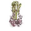
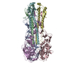
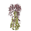
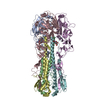
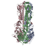
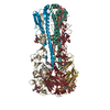
- PDB-3eyk: Structure of Influenza Haemagglutinin in complex with an inhibito... -
+ Open data

- Basic information
| Entry | Database: PDB / ID: 3eyk | ||||||
|---|---|---|---|---|---|---|---|
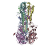
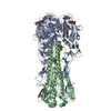
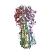
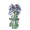
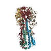
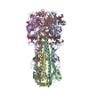
| Title | Structure of Influenza Haemagglutinin in complex with an inhibitor of membrane fusion | ||||||
 Components Components |
| ||||||
 Keywords Keywords |  VIRAL PROTEIN / Influenza / hemagglutinin / inhibitor / Envelope protein / Fusion protein / Glycoprotein / Lipoprotein / Membrane / Palmitate / Transmembrane / Virion VIRAL PROTEIN / Influenza / hemagglutinin / inhibitor / Envelope protein / Fusion protein / Glycoprotein / Lipoprotein / Membrane / Palmitate / Transmembrane / Virion | ||||||
| Function / homology |  Function and homology information Function and homology informationclathrin-dependent endocytosis of virus by host cell / host cell surface receptor binding / fusion of virus membrane with host plasma membrane / fusion of virus membrane with host endosome membrane / viral envelope / virion attachment to host cell / host cell plasma membrane / virion membrane / membraneSimilarity search - Function | ||||||
| Biological species |   Influenza A virus Influenza A virus | ||||||
| Method | X-RAY DIFFRACTION / MOLECULAR REPLACEMENT / Resolution: 2.5 Å | ||||||
 Authors Authors | Russell, R.J. / Kerry, P.S. / Stevens, D.J. / Steinhauer, D.A. / Martin, S.R. / Gamblin, S.J. / Skehel, J.J. | ||||||
 Citation Citation | Journal: Proc.Natl.Acad.Sci.USA / Year: 2008 Title: Structure of influenza hemagglutinin in complex with an inhibitor of membrane fusion Authors: Russell, R.J. / Kerry, P.S. / Stevens, D.J. / Steinhauer, D.A. / Martin, S.R. / Gamblin, S.J. / Skehel, J.J. | ||||||
| History |
|
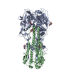
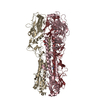
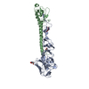
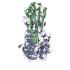
- Structure visualization
| Structure viewer | Molecule: MolmilJmol/JSmol |
|---|
- Downloads & links
-Download
| PDBx/mmCIF format | 3eyk.cif.gz | 116.7 KB | Display | PDBx/mmCIF format |
|---|---|---|---|---|
| PDB format | pdb3eyk.ent.gz | 89.2 KB | Display | PDB format |
| PDBx/mmJSON format | 3eyk.json.gz | Tree view | PDBx/mmJSON format | |
| Others |  Other downloads Other downloads |
-Validation report
| Arichive directory | https://data.pdbj.org/pub/pdb/validation_reports/ey/3eykftp://data.pdbj.org/pub/pdb/validation_reports/ey/3eyk | HTTPS FTP |
|---|
-Related structure data


-Links
 PDBj
PDBj


- Assembly
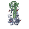
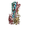
| Deposited unit | 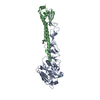
| |||||||||||||||
|---|---|---|---|---|---|---|---|---|---|---|---|---|---|---|---|---|
| 1 | 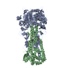
| |||||||||||||||
| Unit cell |
| |||||||||||||||
| Components on special symmetry positions |
|
-Components
| #1: Protein | Mass: 35279.738 Da / Num. of mol.: 1 / Source method: isolated from a natural source / Source: (natural) Influenza A virus / References: UniProt: P26137 |
|---|---|
| #2: Protein | Mass: 19922.895 Da / Num. of mol.: 1 / Source method: isolated from a natural source / Source: (natural) Influenza A virus / References: UniProt: P26137 |
| #3: Chemical | ChemComp-EYK / Tert-Butylhydroquinone  Mass: 166.217 Da / Num. of mol.: 1 / Source method: obtained synthetically / Formula: C10H14O2 Mass: 166.217 Da / Num. of mol.: 1 / Source method: obtained synthetically / Formula: C10H14O2 |
| #4: Water | ChemComp-HOH / Water Mass: 18.015 Da / Num. of mol.: 337 / Source method: isolated from a natural source / Formula: H2O Mass: 18.015 Da / Num. of mol.: 337 / Source method: isolated from a natural source / Formula: H2O |
-Experimental details
-Experiment
| Experiment | Method: X-RAY DIFFRACTION / Number of used crystals: 1 |
|---|
- Sample preparation
| Crystal | Density Matthews: 4.06 Å3/Da / Density % sol: 69.72 % |
|---|---|
| Crystal grow | Temperature: 293 K / Method: vapor diffusion, hanging drop / pH: 8.5 Details: Tris, PEG8000, pH 8.5, VAPOR DIFFUSION, HANGING DROP, temperature 293K |
-Data collection
| Diffraction | Mean temperature: 100 K |
|---|---|
| Diffraction source | Source: ROTATING ANODE / Type: RIGAKU MICROMAX-007 / Wavelength: 1.54 Å |
| Detector | Type: RIGAKU RAXIS IV++ / Detector: IMAGE PLATE / Date: Sep 10, 2007 |
| Radiation | Protocol: SINGLE WAVELENGTH / Monochromatic (M) / Laue (L): M / Scattering type: x-ray |
| Radiation wavelength | Wavelength: 1.54 Å / Relative weight: 1 |
| Reflection | Resolution: 2.5→30 Å / Num. all: 29658 / Num. obs: 28412 / % possible obs: 95.8 % / Observed criterion σ(F): 0 / Observed criterion σ(I): 0 |
- Processing
| Software |
| ||||||||||||||||||||||||||||||||||||||||||||||||||||||||||||||||||||||||||||||||||||||||||||||||||||||||||||||||||||||||||||||||||||||||||||||||||||||||||||||||||||||||||
|---|---|---|---|---|---|---|---|---|---|---|---|---|---|---|---|---|---|---|---|---|---|---|---|---|---|---|---|---|---|---|---|---|---|---|---|---|---|---|---|---|---|---|---|---|---|---|---|---|---|---|---|---|---|---|---|---|---|---|---|---|---|---|---|---|---|---|---|---|---|---|---|---|---|---|---|---|---|---|---|---|---|---|---|---|---|---|---|---|---|---|---|---|---|---|---|---|---|---|---|---|---|---|---|---|---|---|---|---|---|---|---|---|---|---|---|---|---|---|---|---|---|---|---|---|---|---|---|---|---|---|---|---|---|---|---|---|---|---|---|---|---|---|---|---|---|---|---|---|---|---|---|---|---|---|---|---|---|---|---|---|---|---|---|---|---|---|---|---|---|---|---|
| Refinement | Method to determine structure: MOLECULAR REPLACEMENT / Resolution: 2.5→29.66 Å / Cor.coef. Fo:Fc: 0.943 / Cor.coef. Fo:Fc free: 0.912 / SU B: 7.549 / SU ML: 0.166 / Cross valid method: THROUGHOUT / σ(F): 0 / ESU R: 0.307 / ESU R Free: 0.248 / Stereochemistry target values: MAXIMUM LIKELIHOOD / Details: HYDROGENS HAVE BEEN ADDED IN THE RIDING POSITIONS
| ||||||||||||||||||||||||||||||||||||||||||||||||||||||||||||||||||||||||||||||||||||||||||||||||||||||||||||||||||||||||||||||||||||||||||||||||||||||||||||||||||||||||||
| Solvent computation | Ion probe radii: 0.8 Å / Shrinkage radii: 0.8 Å / VDW probe radii: 1.2 Å / Solvent model: MASK | ||||||||||||||||||||||||||||||||||||||||||||||||||||||||||||||||||||||||||||||||||||||||||||||||||||||||||||||||||||||||||||||||||||||||||||||||||||||||||||||||||||||||||
| Displacement parameters | Biso mean: 39.319 Å2 | ||||||||||||||||||||||||||||||||||||||||||||||||||||||||||||||||||||||||||||||||||||||||||||||||||||||||||||||||||||||||||||||||||||||||||||||||||||||||||||||||||||||||||
| Refinement step | Cycle: LAST / Resolution: 2.5→29.66 Å
| ||||||||||||||||||||||||||||||||||||||||||||||||||||||||||||||||||||||||||||||||||||||||||||||||||||||||||||||||||||||||||||||||||||||||||||||||||||||||||||||||||||||||||
| Refine LS restraints |
| ||||||||||||||||||||||||||||||||||||||||||||||||||||||||||||||||||||||||||||||||||||||||||||||||||||||||||||||||||||||||||||||||||||||||||||||||||||||||||||||||||||||||||
| LS refinement shell | Resolution: 2.5→2.566 Å / Total num. of bins used: 20
|
