Resolution: 1.22→41 Å / σ(F): 0 / σ(I): 0 Details: Twinning was observed in the present crystal and refined with SHELXL using the TWIN instruction, resulting in a twin fraction of 0.5, operator h,-h-k,-l. The point group 3 of the crystal ...Details: Twinning was observed in the present crystal and refined with SHELXL using the TWIN instruction, resulting in a twin fraction of 0.5, operator h,-h-k,-l. The point group 3 of the crystal lattice can be obtained via a coset decomposition of point group 32 of the twin lattice. The twofold twin axis cannot be a real crystallographic operation because this would result in only half a molecule in the asymmetric unit.
Rfactor
Num. reflection
% reflection
Rfree
0.222
-
-
Rwork
0.199
-
-
all
-
61949
-
obs
-
61949
97.1 %
Refinement step
Cycle: LAST / Resolution: 1.22→41 Å
Protein
Nucleic acid
Ligand
Solvent
Total
Num. atoms
1430
0
22
73
1525
Refine LS restraints
Refine-ID
Type
Dev ideal
X-RAY DIFFRACTION
s_bond_d
0.02
X-RAY DIFFRACTION
s_angle_d
0.04
+
About Yorodumi
-
News
-
Feb 9, 2022. New format data for meta-information of EMDB entries
New format data for meta-information of EMDB entries
Version 3 of the EMDB header file is now the official format.
The previous official version 1.9 will be removed from the archive.
In the structure databanks used in Yorodumi, some data are registered as the other names, "COVID-19 virus" and "2019-nCoV". Here are the details of the virus and the list of structure data.
Jan 31, 2019. EMDB accession codes are about to change! (news from PDBe EMDB page)
EMDB accession codes are about to change! (news from PDBe EMDB page)
The allocation of 4 digits for EMDB accession codes will soon come to an end. Whilst these codes will remain in use, new EMDB accession codes will include an additional digit and will expand incrementally as the available range of codes is exhausted. The current 4-digit format prefixed with “EMD-” (i.e. EMD-XXXX) will advance to a 5-digit format (i.e. EMD-XXXXX), and so on. It is currently estimated that the 4-digit codes will be depleted around Spring 2019, at which point the 5-digit format will come into force.
The EM Navigator/Yorodumi systems omit the EMD- prefix.
Related info.:Q: What is EMD? / ID/Accession-code notation in Yorodumi/EM Navigator
Yorodumi is a browser for structure data from EMDB, PDB, SASBDB, etc.
This page is also the successor to EM Navigator detail page, and also detail information page/front-end page for Omokage search.
The word "yorodu" (or yorozu) is an old Japanese word meaning "ten thousand". "mi" (miru) is to see.
Related info.:EMDB / PDB / SASBDB / Comparison of 3 databanks / Yorodumi Search / Aug 31, 2016. New EM Navigator & Yorodumi / Yorodumi Papers / Jmol/JSmol / Function and homology information / Changes in new EM Navigator and Yorodumi
 Movie
Movie Controller


 Open data
 Basic information
 Components
 Keywords
 Function and homology information
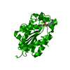
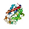
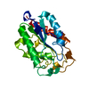
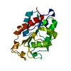
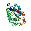
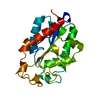
 Fusarium solani f. pisi (fungus)
 Authors
 Citation
 Structure visualization
 Downloads & links
 Other downloads


 PDBj
PDBj
 Assembly


 Mass: 654.871 Da / Num. of mol.: 1 / Source method: obtained synthetically / Formula: C21H27BrNO5PPdS2
 Mass: 18.015 Da / Num. of mol.: 73 / Source method: isolated from a natural source / Formula: H2O
 Sample preparation
 / Beamline: ID29 / Wavelength: 1.0332 Å
 Processing