 Movie
Movie Controller


+ Open data

- Basic information
| Entry | Database: PDB / ID: 1oxm | ||||||
|---|---|---|---|---|---|---|---|
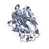
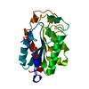
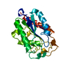
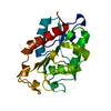
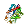
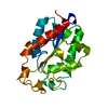
| Title | STRUCTURE OF CUTINASE | ||||||
 Components Components | CUTINASE | ||||||
 Keywords Keywords | SERINE ESTERASE / HYDROLASE / GLYCOPROTEIN | ||||||
| Function / homology |  Function and homology information Function and homology informationcutinase / cutinase activity / carbohydrate catabolic process / extracellular region Similarity search - Function | ||||||
| Biological species |  Nectria haematococca mpVI (fungus) Nectria haematococca mpVI (fungus) | ||||||
| Method |  X-RAY DIFFRACTION / MOLECULAR REPLACEMENT / Resolution: 2.3 Å X-RAY DIFFRACTION / MOLECULAR REPLACEMENT / Resolution: 2.3 Å | ||||||
 Authors Authors | Longhi, S. / Cambillau, C. | ||||||
 Citation Citation | Journal: Protein Sci. / Year: 1997 Title: Crystal structure of cutinase covalently inhibited by a triglyceride analogue. Authors: Longhi, S. / Mannesse, M. / Verheij, H.M. / De Haas, G.H. / Egmond, M. / Knoops-Mouthuy, E. / Cambillau, C. #1: Journal: Proteins / Year: 1996Title: Dynamics of Fusarium Solani Cutinase Investigated Through Structural Comparison Among Different Crystal Forms of its Variants Authors: Longhi, S. / Nicolas, A. / Creveld, L. / Egmond, M. / Verrips, C.T. / De Vlieg, J. / Martinez, C. / Cambillau, C. #2: Journal: Biochemistry / Year: 1996Title: Contribution of Cutinase Serine 42 Side Chain to the Stabilization of the Oxyanion Transition State Authors: Nicolas, A. / Egmond, M. / Verrips, C.T. / De Vlieg, J. / Longhi, S. / Cambillau, C. / Martinez, C. #3: Journal: Biochemistry / Year: 1994Title: Cutinase, a Lipolytic Enzyme with a Preformed Oxyanion Hole Authors: Martinez, C. / Nicolas, A. / Van Tilbeurgh, H. / Egloff, M.P. / Cudrey, C. / Verger, R. / Cambillau, C. #4: Journal: Nature / Year: 1992Title: Fusarium Solani Cutinase is a Lipolytic Enzyme with a Catalytic Serine Accessible to Solvent Authors: Martinez, C. / De Geus, P. / Lauwereys, M. / Matthyssens, G. / Cambillau, C. | ||||||
| History |
|
- Structure visualization
| Structure viewer | Molecule: MolmilJmol/JSmol |
|---|
- Downloads & links
-Download
| PDBx/mmCIF format | 1oxm.cif.gz | 111.9 KB | Display | PDBx/mmCIF format |
|---|---|---|---|---|
| PDB format | pdb1oxm.ent.gz | 86.7 KB | Display | PDB format |
| PDBx/mmJSON format | 1oxm.json.gz | Tree view | PDBx/mmJSON format | |
| Others |  Other downloads Other downloads |
-Validation report
| Arichive directory | https://data.pdbj.org/pub/pdb/validation_reports/ox/1oxmftp://data.pdbj.org/pub/pdb/validation_reports/ox/1oxm | HTTPS FTP |
|---|
-Related structure data
| Related structure data | 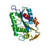 1cusS S: Starting model for refinement |
|---|---|
| Similar structure data |


-Links
 PDBj
PDBj
- Assembly
| Deposited unit | 
| ||||||||
|---|---|---|---|---|---|---|---|---|---|
| 1 | 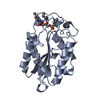
| ||||||||
| 2 | 
| ||||||||
| Unit cell |
| ||||||||
| Noncrystallographic symmetry (NCS) | NCS oper: (Code: given Matrix: (-0.62389, -0.46433, -0.62862), Vector: Details | THERE ARE TWO MOLECULES PER ASYMMETRIC UNIT. | |
-Components
| #1: Protein | Mass: 22278.953 Da / Num. of mol.: 2 Source method: isolated from a genetically manipulated source Source: (gene. exp.) Nectria haematococca mpVI (fungus) / Species: Nectria haematococca / Strain: mpVI / Plasmid: MIRY / Production host:  References: UniProt: P00590, Hydrolases; Acting on ester bonds; Carboxylic-ester hydrolases #2: Chemical |   Mass: 394.443 Da / Num. of mol.: 2 / Source method: obtained synthetically / Formula: C17H35N2O6P Mass: 394.443 Da / Num. of mol.: 2 / Source method: obtained synthetically / Formula: C17H35N2O6P#3: Water | ChemComp-HOH / |  Mass: 18.015 Da / Num. of mol.: 209 / Source method: isolated from a natural source / Formula: H2O Mass: 18.015 Da / Num. of mol.: 209 / Source method: isolated from a natural source / Formula: H2OCompound details | WT CUTINASE COVALENTLY INHIBITED BY THE TRIGLYCERIDE ANALOGUE TC4 BOUND TO THE CATALYTIC SERINE ...WT CUTINASE COVALENTLY | Has protein modification | Y | |
|---|
-Experimental details
-Experiment
| Experiment | Method: X-RAY DIFFRACTION / Number of used crystals: 1 |
|---|
- Sample preparation
| Crystal | Density Matthews: 2.11 Å3/Da / Density % sol: 42 % | ||||||||||||||||||||||||||||||
|---|---|---|---|---|---|---|---|---|---|---|---|---|---|---|---|---|---|---|---|---|---|---|---|---|---|---|---|---|---|---|---|
| Crystal grow | pH: 7 / Details: PEG 6000 IN HEPES 0.1 M PH 7, pH 7.0 | ||||||||||||||||||||||||||||||
| Crystal grow | *PLUS Temperature: 20 ℃ / Method: vapor diffusion, hanging drop / Details: Abergel, C., (1990) J. Mol. Biol., 215, 215. | ||||||||||||||||||||||||||||||
| Components of the solutions | *PLUS
|
-Data collection
| Diffraction | Mean temperature: 291 K |
|---|---|
| Diffraction source | Wavelength: 1.5418 |
| Detector | Type: MARRESEARCH / Detector: IMAGE PLATE |
| Radiation | Monochromatic (M) / Laue (L): M / Scattering type: x-ray |
| Radiation wavelength | Wavelength: 1.5418 Å / Relative weight: 1 |
| Reflection | Resolution: 2.3→30 Å / Num. obs: 15569 / % possible obs: 93.8 % / Observed criterion σ(I): 0 / Redundancy: 2.94 % / Rmerge(I) obs: 0.0523 / Net I/σ(I): 25.13 |
| Reflection shell | Resolution: 2.3→2.4 Å / Redundancy: 2.3 % / Rmerge(I) obs: 0.1768 / Mean I/σ(I) obs: 6.9 / % possible all: 67.7 |
| Reflection | *PLUS Num. measured all: 45670 |
- Processing
| Software |
| ||||||||||||||||||||||||||||||||||||||||||||||||||||||||||||
|---|---|---|---|---|---|---|---|---|---|---|---|---|---|---|---|---|---|---|---|---|---|---|---|---|---|---|---|---|---|---|---|---|---|---|---|---|---|---|---|---|---|---|---|---|---|---|---|---|---|---|---|---|---|---|---|---|---|---|---|---|---|
| Refinement | Method to determine structure: MOLECULAR REPLACEMENT Starting model: NATIVE STRUCTURE AT 1.6 ANGSTROMS (PDB ENTRY 1CUS) Resolution: 2.3→30 Å / Cross valid method: R-FREE / σ(F): 0 Details: THE CATALYTIC SER 120 OF BOTH MOLECULES IN THE ASYMMETRIC UNIT LIES IN AN UNFAVORABLE REGION OF THE RAMACHANDRAN PLOT (EPSILON CONFORMATION) WHICH IS TYPICAL OF ALL MEMBERS OF THE ALPHA/BETA ...Details: THE CATALYTIC SER 120 OF BOTH MOLECULES IN THE ASYMMETRIC UNIT LIES IN AN UNFAVORABLE REGION OF THE RAMACHANDRAN PLOT (EPSILON CONFORMATION) WHICH IS TYPICAL OF ALL MEMBERS OF THE ALPHA/BETA HYDROLASE SUPERFAMILY.
| ||||||||||||||||||||||||||||||||||||||||||||||||||||||||||||
| Refine analyze | Luzzati coordinate error obs: 0.15 Å / Luzzati sigma a obs: 0.28 Å | ||||||||||||||||||||||||||||||||||||||||||||||||||||||||||||
| Refinement step | Cycle: LAST / Resolution: 2.3→30 Å
| ||||||||||||||||||||||||||||||||||||||||||||||||||||||||||||
| Refine LS restraints |
|
