 Movie
Movie Controller


+ Open data

- Basic information
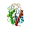
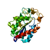
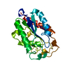
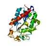
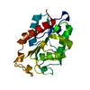
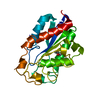
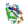
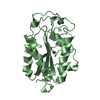
| Entry | Database: PDB / ID: 1cex | ||||||
|---|---|---|---|---|---|---|---|
| Title | STRUCTURE OF CUTINASE | ||||||
 Components Components | CUTINASE | ||||||
 Keywords Keywords | SERINE ESTERASE / HYDROLASE / GLYCOPROTEIN | ||||||
| Function / homology |  Function and homology information Function and homology informationcutinase / cutinase activity / carbohydrate catabolic process / extracellular region Similarity search - Function | ||||||
| Biological species |  Nectria haematococca mpVI (fungus) Nectria haematococca mpVI (fungus) | ||||||
| Method |  X-RAY DIFFRACTION / SYNCHROTRON / Resolution: 1 Å X-RAY DIFFRACTION / SYNCHROTRON / Resolution: 1 Å | ||||||
 Authors Authors | Longhi, S. / Czjzek, M. / Lamzin, V. / Nicolas, A. / Cambillau, C. | ||||||
 Citation Citation | Journal: J.Mol.Biol. / Year: 1997 Title: Atomic resolution (1.0 A) crystal structure of Fusarium solani cutinase: stereochemical analysis. Authors: Longhi, S. / Czjzek, M. / Lamzin, V. / Nicolas, A. / Cambillau, C. #1: Journal: Protein Sci. / Year: 1997Title: Crystal Structure of Cutinase Covalently Inhibited by a Triglyceride Analogue Authors: Longhi, S. / Mannesse, M. / Verheij, H.M. / De Haas, G.H. / Egmond, M. / Knoops-Mouthuy, E. / Cambillau, C. #2: Journal: Proteins / Year: 1996Title: Dynamics of Fusarium Solani Cutinase Investigated Through Structural Comparison Among Different Crystal Forms of its Variants Authors: Longhi, S. / Nicolas, A. / Creveld, L. / Egmond, M. / Verrips, C.T. / De Vlieg, J. / Martinez, C. / Cambillau, C. #3: Journal: Biochemistry / Year: 1996Title: Contribution of Cutinase Serine 42 Side Chain to the Stabilization of the Oxyanion Transition State Authors: Nicolas, A. / Egmond, M. / Verrips, C.T. / De Vlieg, J. / Longhi, S. / Cambillau, C. / Martinez, C. #4: Journal: Biochemistry / Year: 1994Title: Cutinase, a Lipolytic Enzyme with a Preformed Oxyanion Hole Authors: Martinez, C. / Nicolas, A. / Van Tilbeurgh, H. / Egloff, M.P. / Cudrey, C. / Verger, R. / Cambillau, C. #5: Journal: Nature / Year: 1992Title: Fusarium Solani Cutinase is a Lipolytic Enzyme with a Catalytic Serine Accessible to Solvent Authors: Martinez, C. / De Geus, P. / Lauwereys, M. / Matthyssens, G. / Cambillau, C. | ||||||
| History |
|
- Structure visualization
| Structure viewer | Molecule: MolmilJmol/JSmol |
|---|
- Downloads & links
-Download
| PDBx/mmCIF format | 1cex.cif.gz | 87.2 KB | Display | PDBx/mmCIF format |
|---|---|---|---|---|
| PDB format | pdb1cex.ent.gz | 67.3 KB | Display | PDB format |
| PDBx/mmJSON format | 1cex.json.gz | Tree view | PDBx/mmJSON format | |
| Others |  Other downloads Other downloads |
-Validation report
| Arichive directory | https://data.pdbj.org/pub/pdb/validation_reports/ce/1cexftp://data.pdbj.org/pub/pdb/validation_reports/ce/1cex | HTTPS FTP |
|---|
-Related structure data


-Links
 PDBj
PDBj
- Assembly
| Deposited unit | 
| ||||||||
|---|---|---|---|---|---|---|---|---|---|
| 1 |
| ||||||||
| Unit cell |
|
-Components
| #1: Protein | Mass: 22278.953 Da / Num. of mol.: 1 Source method: isolated from a genetically manipulated source Source: (gene. exp.) Nectria haematococca mpVI (fungus) / Species: Nectria haematococca / Strain: mpVI / Plasmid: MIRY / Production host:  References: UniProt: P00590, Hydrolases; Acting on ester bonds; Carboxylic-ester hydrolases |
|---|---|
| #2: Water | ChemComp-HOH /  Mass: 18.015 Da / Num. of mol.: 264 / Source method: isolated from a natural source / Formula: H2O Mass: 18.015 Da / Num. of mol.: 264 / Source method: isolated from a natural source / Formula: H2O |
| Compound details | THE CATALYTIC SERINE HAS THE EPSILON CONFORMATION WHICH IS TYPICAL OF ALL THE MEMBERS OF THE ...THE CATALYTIC SERINE HAS THE EPSILON CONFORMATI |
| Has protein modification | Y |
-Experimental details
-Experiment
| Experiment | Method: X-RAY DIFFRACTION |
|---|
- Sample preparation
| Crystal | Density Matthews: 1.96 Å3/Da / Density % sol: 37 % | ||||||||||||||||||||
|---|---|---|---|---|---|---|---|---|---|---|---|---|---|---|---|---|---|---|---|---|---|
| Crystal grow | *PLUS Temperature: 20 ℃ / pH: 7 / Method: vapor diffusion, hanging drop / Details: Abergel, C., (1990) J. Mol. Biol., 215, 215. | ||||||||||||||||||||
| Components of the solutions | *PLUS
|
-Data collection
| Diffraction source | Source: SYNCHROTRON / Site: EMBL/DESY, HAMBURG  / Beamline: X11 / Wavelength: 0.927 / Beamline: X11 / Wavelength: 0.927 |
|---|---|
| Detector | Type: MAR scanner 300 mm plate / Detector: IMAGE PLATE |
| Radiation | Monochromatic (M) / Laue (L): M / Scattering type: x-ray |
| Radiation wavelength | Wavelength: 0.927 Å / Relative weight: 1 |
| Reflection | Num. obs: 86474 / % possible obs: 93.3 % / Redundancy: 2.07 % / Rmerge(I) obs: 0.039 |
| Reflection | *PLUS Highest resolution: 1 Å / Lowest resolution: 15 Å / Num. measured all: 179064 |
| Reflection shell | *PLUS Highest resolution: 1 Å / Lowest resolution: 1.01 Å / % possible obs: 99 % / Rmerge(I) obs: 0.598 / Mean I/σ(I) obs: 2.2 |
- Processing
| Software |
| |||||||||||||||||||||||||||||||||
|---|---|---|---|---|---|---|---|---|---|---|---|---|---|---|---|---|---|---|---|---|---|---|---|---|---|---|---|---|---|---|---|---|---|---|
| Refinement | Resolution: 1→15 Å / σ(F): 0 / Stereochemistry target values: ENGH & HUBER
| |||||||||||||||||||||||||||||||||
| Refinement step | Cycle: LAST / Resolution: 1→15 Å
| |||||||||||||||||||||||||||||||||
| Refine LS restraints |
|
