 Movie
Movie Controller

[English] 日本語
 Yorodumi
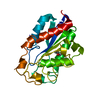
- PDB-1agy: The 1.15 angstrom refined structure of fusarium solani pisi cutinase -
+ Open data

- Basic information
| Entry | Database: PDB / ID: 1agy | ||||||
|---|---|---|---|---|---|---|---|
| Title | The 1.15 angstrom refined structure of fusarium solani pisi cutinase | ||||||
 Components Components | CUTINASE | ||||||
 Keywords Keywords | SERINE ESTERASE / HYDROLASE / GLYCOPROTEIN | ||||||
| Function / homology |  Function and homology information Function and homology informationcutinase / cutinase activity / carbohydrate catabolic process / extracellular region Similarity search - Function | ||||||
| Biological species |  Nectria haematococca mpVI (fungus) Nectria haematococca mpVI (fungus) | ||||||
| Method |  X-RAY DIFFRACTION / RESOLUTION EXTENSION / Resolution: 1.15 Å X-RAY DIFFRACTION / RESOLUTION EXTENSION / Resolution: 1.15 Å | ||||||
 Authors Authors | Nicolas, A. / Martinez, C. / Cambillau, C. | ||||||
 Citation Citation | Journal: J.Mol.Biol. / Year: 1997 Title: Atomic resolution (1.0 A) crystal structure of Fusarium solani cutinase: stereochemical analysis. Authors: Longhi, S. / Czjzek, M. / Lamzin, V. / Nicolas, A. / Cambillau, C. #1: Journal: Nature / Year: 1992Title: Fusarium Solani Cutinase is a Lipolytic Enzyme with a Catalytic Serine Accessible to Solvent Authors: Martinez, C. / De Geus, P. / Lauwereys, M. / Matthyssens, G. / Cambillau, C. | ||||||
| History |
|
- Structure visualization
| Structure viewer | Molecule: MolmilJmol/JSmol |
|---|
- Downloads & links
-Download
| PDBx/mmCIF format | 1agy.cif.gz | 105.3 KB | Display | PDBx/mmCIF format |
|---|---|---|---|---|
| PDB format | pdb1agy.ent.gz | 84 KB | Display | PDB format |
| PDBx/mmJSON format | 1agy.json.gz | Tree view | PDBx/mmJSON format | |
| Others |  Other downloads Other downloads |
-Validation report
| Arichive directory | https://data.pdbj.org/pub/pdb/validation_reports/ag/1agyftp://data.pdbj.org/pub/pdb/validation_reports/ag/1agy | HTTPS FTP |
|---|
-Related structure data


-Links
 PDBj
PDBj
- Assembly
| Deposited unit | 
| ||||||||
|---|---|---|---|---|---|---|---|---|---|
| 1 |
| ||||||||
| Unit cell |
|
-Components
| #1: Protein | Mass: 20828.400 Da / Num. of mol.: 1 Source method: isolated from a genetically manipulated source Source: (gene. exp.) Nectria haematococca mpVI (fungus) / Species: Nectria haematococca / Strain: mpVI / Production host:  References: UniProt: P00590, Hydrolases; Acting on ester bonds; Carboxylic-ester hydrolases |
|---|---|
| #2: Water | ChemComp-HOH /  Mass: 18.015 Da / Num. of mol.: 270 / Source method: isolated from a natural source / Formula: H2O Mass: 18.015 Da / Num. of mol.: 270 / Source method: isolated from a natural source / Formula: H2O |
| Has protein modification | Y |
| Sequence details | REGARDING IDENTITY OF RESIDUE ARG 32, UPON QUESTIONING OF TOMMY CARSTENSEN, AUTHORS SONIA LONGHI ...REGARDING IDENTITY OF RESIDUE ARG 32, UPON QUESTIONIN |
-Experimental details
-Experiment
| Experiment | Method: X-RAY DIFFRACTION / Number of used crystals: 2 |
|---|
- Sample preparation
| Crystal | Density Matthews: 1.98 Å3/Da / Density % sol: 38 % | ||||||||||||||||||||
|---|---|---|---|---|---|---|---|---|---|---|---|---|---|---|---|---|---|---|---|---|---|
| Crystal grow | pH: 7 Details: PROTEIN WAS CRYSTALLIZED FROM 15 - 20% PEG 6000, 0.1 M HEPES, PH 7.0 | ||||||||||||||||||||
| Crystal grow | *PLUS Temperature: 20 ℃ / Method: vapor diffusion, hanging drop / Details: Abergel, C., (1990) J. Mol. Biol., 215, 215. | ||||||||||||||||||||
| Components of the solutions | *PLUS
|
-Data collection
| Diffraction | Mean temperature: 288 K |
|---|---|
| Diffraction source | Source: ROTATING ANODE / Type: RIGAKU RUH2R / Wavelength: 1.5418 |
| Detector | Type: NICOLET / Detector: AREA DETECTOR / Date: Jan 1, 1993 / Details: NO |
| Radiation | Monochromator: SI(111) / Monochromatic (M) / Laue (L): M / Scattering type: x-ray |
| Radiation wavelength | Wavelength: 1.5418 Å / Relative weight: 1 |
| Reflection | Resolution: 1.15→20 Å / Num. obs: 60972 / % possible obs: 95.22 % / Observed criterion σ(I): 0 / Redundancy: 4.09 % / Biso Wilson estimate: 8.43 Å2 / Rmerge(I) obs: 0.01 / Rsym value: 0.01 / Net I/σ(I): 27.46 |
| Reflection shell | Resolution: 1.15→1.2 Å / Redundancy: 2 % / Rmerge(I) obs: 0.03 / Mean I/σ(I) obs: 3.21 / Rsym value: 0.03 / % possible all: 84.2 |
- Processing
| Software |
| ||||||||||||||||||||||||||||||||||||||||||||||||||||||||||||
|---|---|---|---|---|---|---|---|---|---|---|---|---|---|---|---|---|---|---|---|---|---|---|---|---|---|---|---|---|---|---|---|---|---|---|---|---|---|---|---|---|---|---|---|---|---|---|---|---|---|---|---|---|---|---|---|---|---|---|---|---|---|
| Refinement | Method to determine structure: RESOLUTION EXTENSION Starting model: CUTINASE STRUCTURE AT 1.6 ANGSTROM RESOLUTION Resolution: 1.15→20 Å / Rfactor Rfree error: 0 / Data cutoff high absF: 1.15 / Data cutoff low absF: 6 / σ(F): 0
| ||||||||||||||||||||||||||||||||||||||||||||||||||||||||||||
| Displacement parameters | Biso mean: 13.54 Å2 | ||||||||||||||||||||||||||||||||||||||||||||||||||||||||||||
| Refine analyze | Luzzati sigma a obs: 0.01 Å | ||||||||||||||||||||||||||||||||||||||||||||||||||||||||||||
| Refinement step | Cycle: LAST / Resolution: 1.15→20 Å
| ||||||||||||||||||||||||||||||||||||||||||||||||||||||||||||
| Refine LS restraints |
| ||||||||||||||||||||||||||||||||||||||||||||||||||||||||||||
| LS refinement shell | Resolution: 1.15→1.2 Å / Rfactor Rfree error: 0 / Total num. of bins used: 8
| ||||||||||||||||||||||||||||||||||||||||||||||||||||||||||||
| Xplor file |
| ||||||||||||||||||||||||||||||||||||||||||||||||||||||||||||
| Software | *PLUS Name: X-PLOR / Version: 3.1 / Classification: refinement | ||||||||||||||||||||||||||||||||||||||||||||||||||||||||||||
| Refine LS restraints | *PLUS
| ||||||||||||||||||||||||||||||||||||||||||||||||||||||||||||
| LS refinement shell | *PLUS Rfactor Rfree: 0.4 / Rfactor Rwork: 0.4 |
