- PDB-3dep: Structural basis for specific substrate recognition by the chloro... -
+
Open data
ID or keywords:
Loading...
-
Basic information
Entry
Database: PDB / ID: 3dep
Title
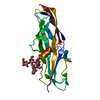
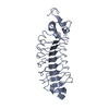
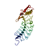
Structural basis for specific substrate recognition by the chloroplast signal recognition particle protein cpSRP43
Components
Signal recognition particle 43 kDa protein
YPGGSFDPLGLA
Keywords
PROTEIN TRANSPORT / MEMBRANE PROTEIN / Chloroplast SRP system / Signal recognition particle / signal sequence / ankyrin repeat / chromodomain / type I turn / substrate protein recognition / L18 region / LHCP / ANK repeat / Chloroplast / Coiled coil / Plastid / Ribonucleoprotein
Function / homology
Function and homology information
protein import into chloroplast thylakoid membrane / protein heterotrimerization / signal recognition particle, endoplasmic reticulum targeting / response to high light intensity / chloroplast envelope / chloroplast stroma / chloroplast thylakoid membrane / chloroplast / disordered domain specific binding / protein-macromolecule adaptor activity ...protein import into chloroplast thylakoid membrane / protein heterotrimerization / signal recognition particle, endoplasmic reticulum targeting / response to high light intensity / chloroplast envelope / chloroplast stroma / chloroplast thylakoid membrane / chloroplast / disordered domain specific binding / protein-macromolecule adaptor activity / protein domain specific binding / protein-containing complex / metal ion binding / identical protein binding / cytosol Similarity search - Function
In the structure databanks used in Yorodumi, some data are registered as the other names, "COVID-19 virus" and "2019-nCoV". Here are the details of the virus and the list of structure data.
Jan 31, 2019. EMDB accession codes are about to change! (news from PDBe EMDB page)
EMDB accession codes are about to change! (news from PDBe EMDB page)
The allocation of 4 digits for EMDB accession codes will soon come to an end. Whilst these codes will remain in use, new EMDB accession codes will include an additional digit and will expand incrementally as the available range of codes is exhausted. The current 4-digit format prefixed with “EMD-” (i.e. EMD-XXXX) will advance to a 5-digit format (i.e. EMD-XXXXX), and so on. It is currently estimated that the 4-digit codes will be depleted around Spring 2019, at which point the 5-digit format will come into force.
The EM Navigator/Yorodumi systems omit the EMD- prefix.
Related info.:Q: What is EMD? / ID/Accession-code notation in Yorodumi/EM Navigator
Yorodumi is a browser for structure data from EMDB, PDB, SASBDB, etc.
This page is also the successor to EM Navigator detail page, and also detail information page/front-end page for Omokage search.
The word "yorodu" (or yorozu) is an old Japanese word meaning "ten thousand". "mi" (miru) is to see.
Related info.:EMDB / PDB / SASBDB / Comparison of 3 databanks / Yorodumi Search / Aug 31, 2016. New EM Navigator & Yorodumi / Yorodumi Papers / Jmol/JSmol / Function and homology information / Changes in new EM Navigator and Yorodumi
 Movie
Movie Controller

 Yorodumi
 Open data
 Basic information
 Components
 Keywords
 Function and homology information
 X-RAY DIFFRACTION /
  Authors
 Citation
 Structure visualization
 Downloads & links
 Other downloads
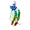


 PDBj
PDBj

 Assembly


 Mass: 35.453 Da / Num. of mol.: 1 / Source method: obtained synthetically / Formula: Cl
 Mass: 18.015 Da / Num. of mol.: 8 / Source method: isolated from a natural source / Formula: H2O
 Sample preparation
 / Beamline: ID14-3 / Wavelength: 0.931 Å
 Processing